Illustrations
Illustration carpal tunnel syndrome
Illustration of the causes of carpal tunnel syndrome, i.e., impairment of the median nerve due to swelling or inflammation at the carpal tunnel.
Illustration of the causes of carpal tunnel syndrome, i.e., impairment of the median nerve due to swelling or inflammation at the carpal tunnel.
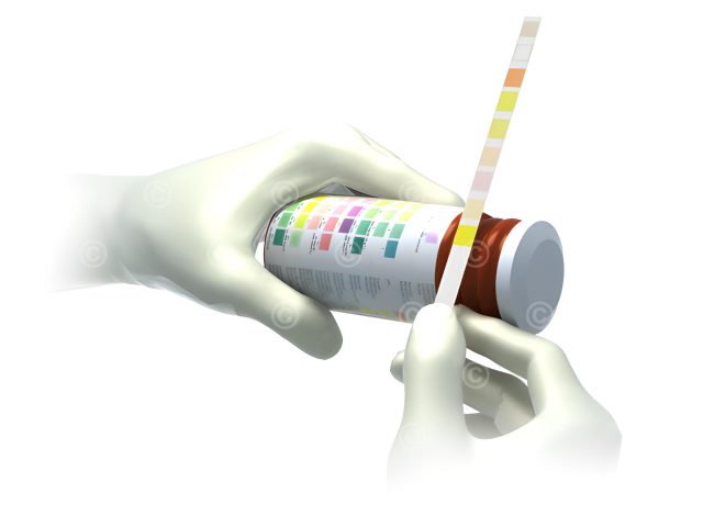
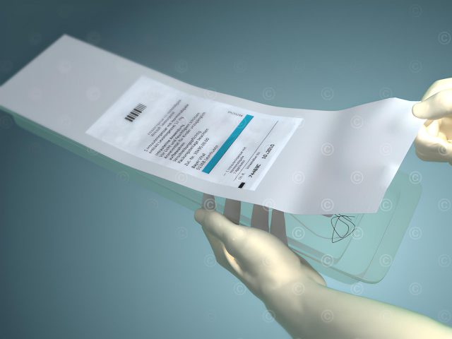
Illustration of a rapid urine test using urine test strips.

Illustration of a Pap smear in sectional view and a histological section of the cells of the of the cervix.

Illustration “anatomy of the spine”. Illustration of the sections of the spinal column and the structure of vertebral segments and discs.

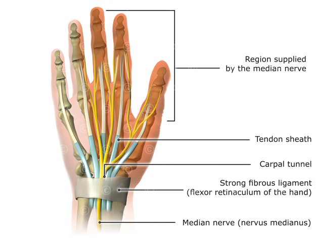
Illustration of the anatomy of the human hand with skeleton, vessels, nerves, ligaments and tendons.
Illustration of a uterine cell compared to cell transformed by HPV (human papillom viruses).
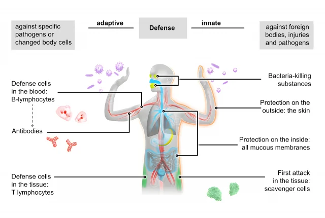
Medical illustrations of the two parts of the immune system: innate general (unspecific) defense and acquired specialized (adaptive) defense.

Illustration of the anatomy of the kidney in with ureter, kidney capsule, kidney cortex, kidney, renal vein and renal artery.

Schematic illustration of the anatomy of a joint showing the joint capsule, articular gap, articular cartilage, bones and muscles.
3D animation about the “application and mode-of-action of a contraceptive loop”.
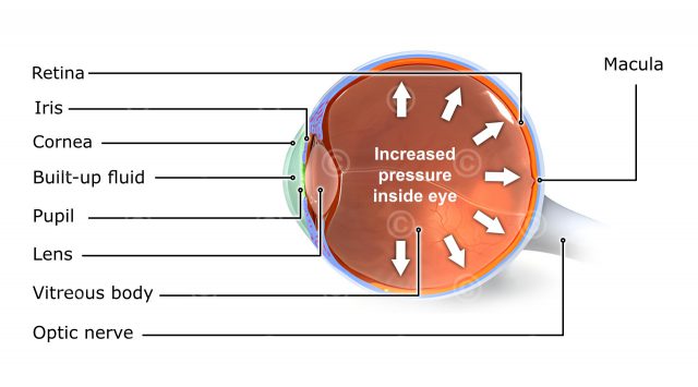
Illustration of the anatomy of the eye and the role of elevated intraocular pressure for the development of a glaucoma.
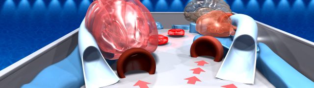
Content for serious game pinball game for a booth at a medical convention (implemented with Unity) and trailer animation.
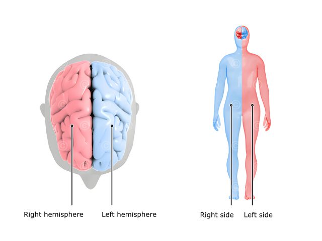
Illustrations about the competence of one half of the brain to the other half of the body (crossover).
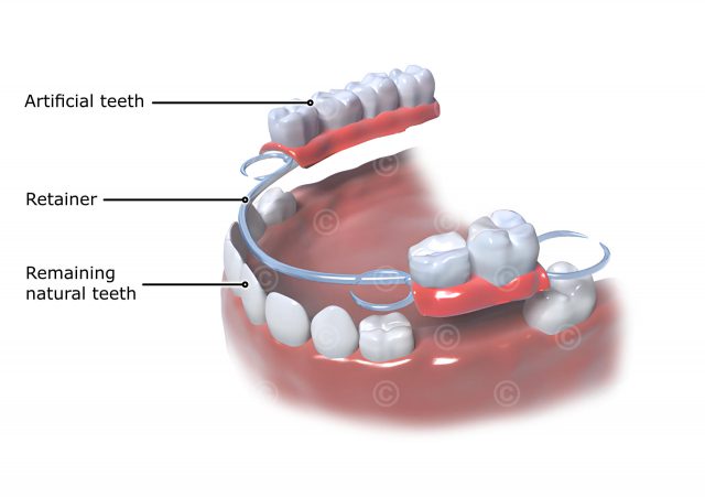
Illustrations of a dental prosthesis using the example of a partial denture on the lower jaw (clasp denture).
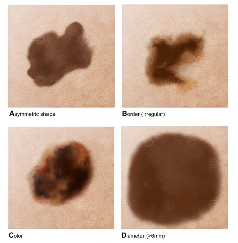
Illustrations on the criterias for the detection of demal changes in a skin cancer (melanoma).

Illustrations on the anatomy of the brain in with the cerebrum, phlegm, thalamus, hypothalamus, hypophysis, cerebellum.
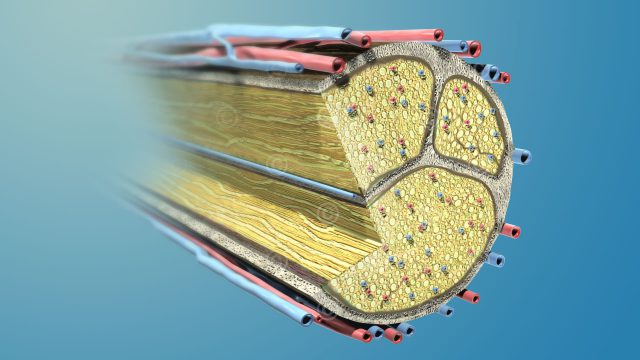
Illustrations of the nervous system. Illustration of a nerve with nerve fiber bundle and supplying blood vessels.
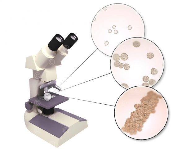
Illustration of a microscope including microscopic pictures of propagating bacteria.
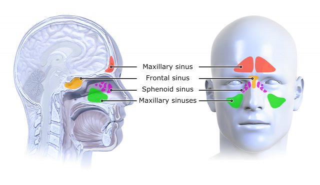
Medical illustrations of paranasal sinuses and their position in the body.
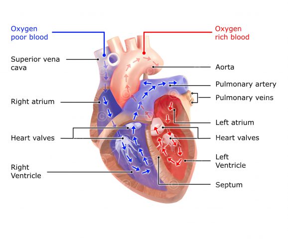
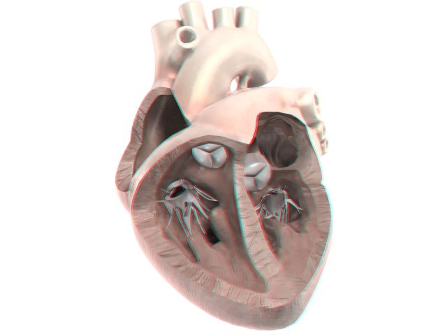
Illustration about of blood circulation of the two blood circuits of the heart. Illustration of the heart in the 4-chamber view (atrium and ventricle).
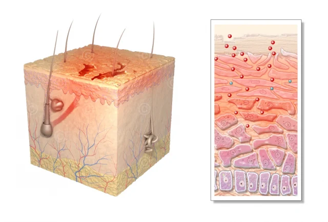
Illustrations of macroscopic and histologic changes of the skin in atopic dermatitis.
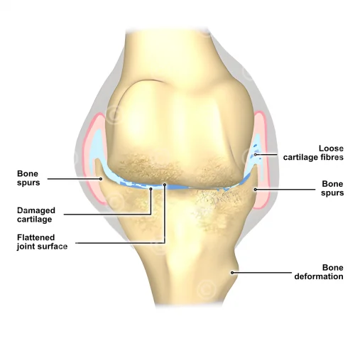
Illustration of incipient osteoarthritis of the knee joint and an illustration of advanced osteoarthritis of the knee.
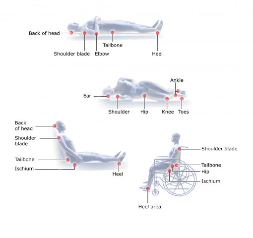
Illustrations about the most affected areas of bedsore.
Stereoscopic 3D film about fat metabolism of the body using anaglyph technique (red cyan glasses)
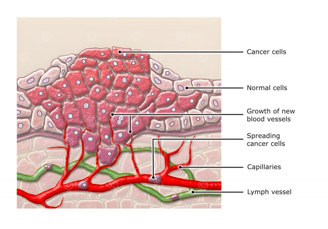
Histological illustrations of healthy cells and cell division, tumor formation and tumor metastasis.
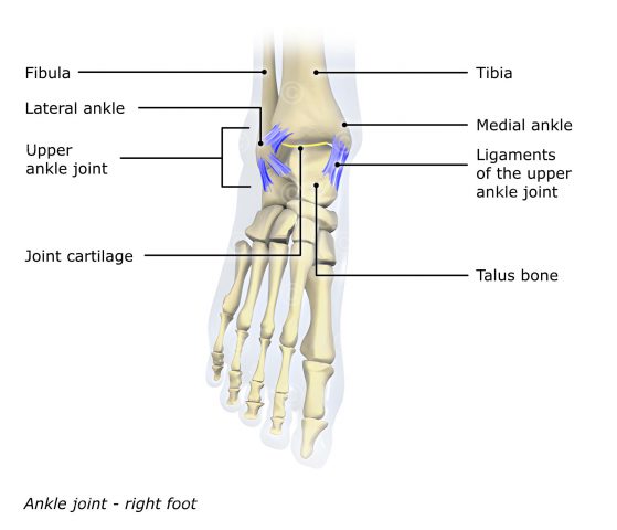
Anatomische Illustrationen zu den Bändern und der Postion des Sprunggelenks.

Project description of an animation for an information campaign covering hyperthyroidism for TV.
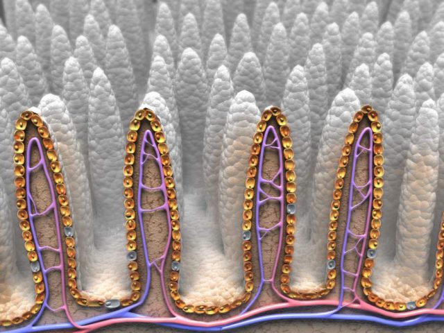
Illustration of healthy intestinal villi in a sectional view with epithelial cells and blood vessels.
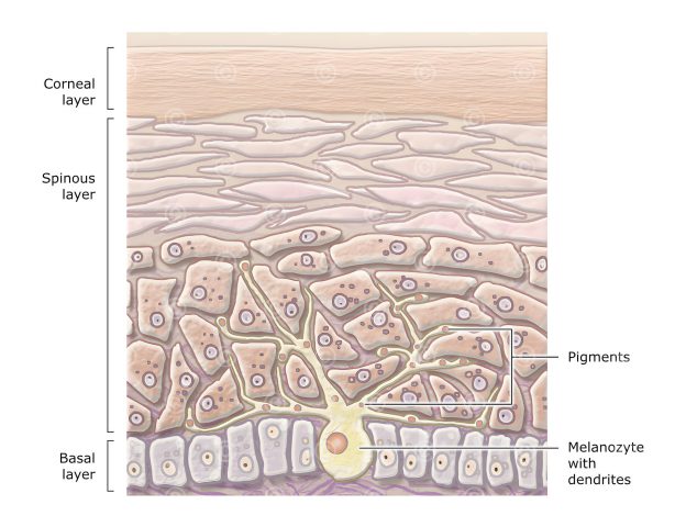
Medical illustration of a melanocyte (pigment cell) in the basal layer (stratum basale) and the spinal layer (stratum spinosum) of the skin.
