Design of an illustration of healthy heart excitation compared to conduction in a heart with atrial fibrillation.
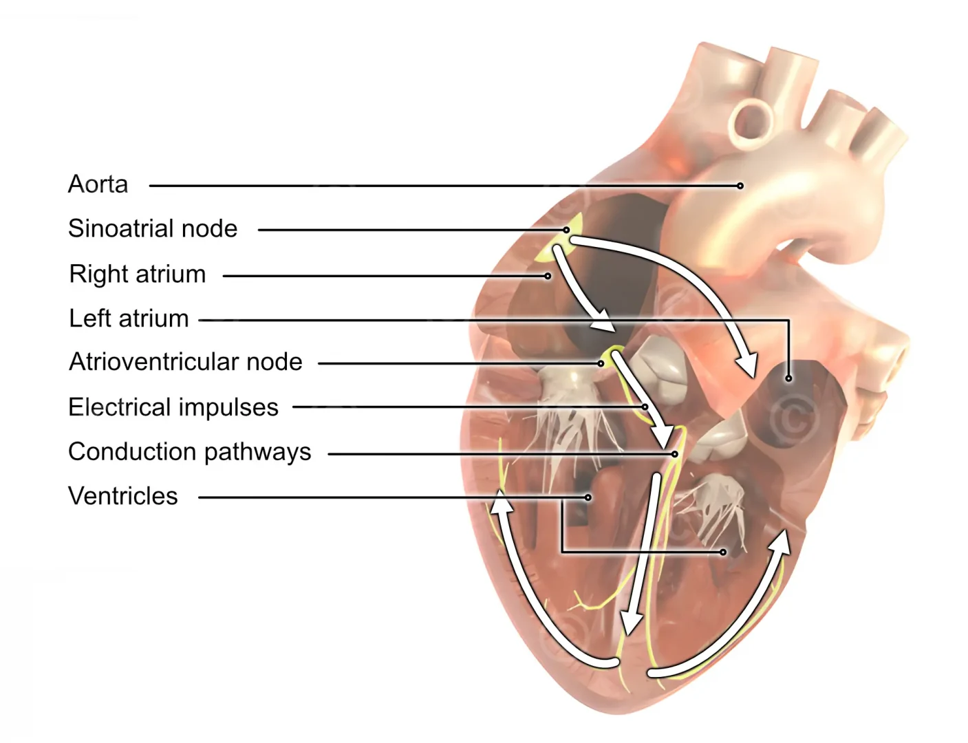
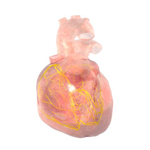
Cardiac conduction is an autonomous process that enables the rhythmic contraction of the heart muscle. This cardiac excitation begins in the sinus node, which is located in the right atrium of the heart and consists of specialized cardiac muscle cells. The sinus node generates electrical impulses that trigger the contraction of the heart and thus determine the heart rhythm.
These impulses propagate through the right atrium and stimulate contraction of the atria, which pumps blood into the ventricles.
From the atria, the impulses travel to the AV node (atrioventricular node), a region between the atria and the ventricles. The AV node slows the electrical impulses to give the ventricles enough time to fill with blood before they then contract.
Next, the impulses propagate through the His bundle and the Tawara legs in the septum via the Purkinje fibers in the ventricles. This causes the ventricles (heart chambers) to contract and blood is pumped into the major arteries (pulmonary circulation, systemic circulation).
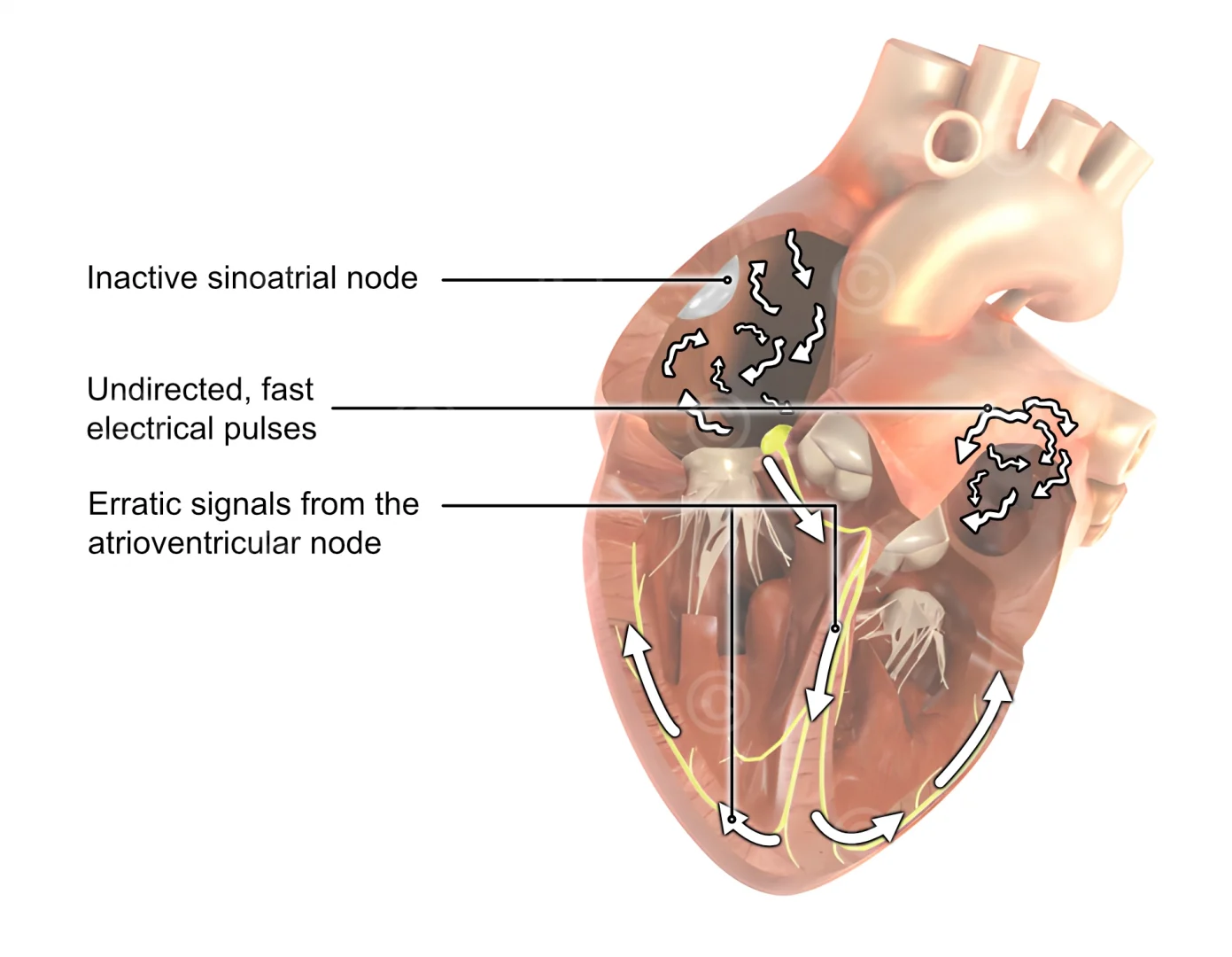
In atrial fibrillation, undirected electrical excitations run across the atria, triggering insufficient contraction of the atria. The atria pump irregularly and very rapidly, impairing the effective pumping function of the entire heart.
Content: Illustration atrial fibrillation
Utilization: Patient information website
Specifications: 640 * 480 pixel
Client: Dimensional / Gesundheitsinformation.de – IQWIG
The rights of use of the illustrations shown are with the respective clients. The shown images are protected with watermarks.
