Description
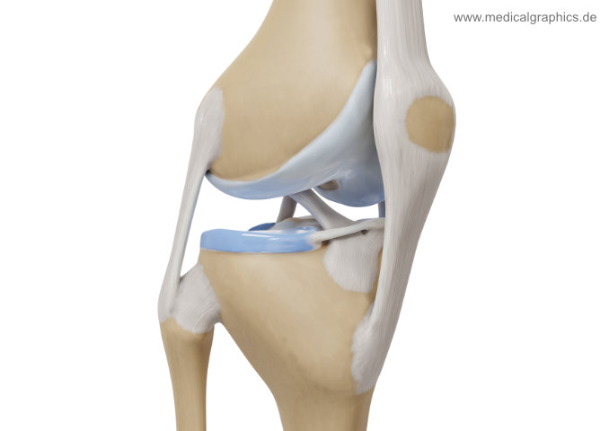
Free illustration of the anatomy of the human knee with ligaments in a right front view. Visible are the anterior and posterior cruciate ligaments, the lateral collateral ligament, the external mesicus, femur, tibia and fibula, patella and patellar tendon.
Note: In this picture the distance from femur to tibia/fibula has been for better visibility of the menisci and ligaments.
Keywords: anatomy, anterior cruciate ligament, ACL, posterior cruciate ligament, PCL, medial collateral ligament, MCL, lateral collateral ligament, LCL, femur, fibula, illustration, internal ligament, knee, ligaments, menisci, patella, patella tendon, quadriceps tendon, tibia, front side, lateral anterior, free, free image, creative commons license, cc