PD-1 binds to PD-L1 – dark

Free illustration of the binding of the surface protein PD-L1 to PD-1 as a surface model with indicated ribbon model on a dark background. Programmed cell death 1 ligand 1 is involved in the inhibition of the immune response together with the receptor PD-1. PD-1 (red) is found on the surface of T cells and interacts with the ligand protein PD-L1 which is found on the surface of regulatory cells, such as the cells that present antigens from invaders. – Illustration based on structural data from Lin, D.Y., Tanaka, Y., Iwasaki, M., Gittis, A.G., Su, H.P., Mikami, B., Okazaki, T., Honjo, T., Minato, N., Garboczi, D.N.

Description

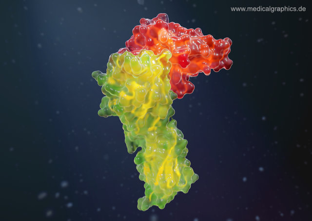
Information illustration – Title – PD-1 binds to PD-L1 – dark – Keywords – free, illustration, binding, surface protein, PD-L1, PD-1, surface model, ribbon model, dark background, programmed cell death 1 ligand 1, inhibition, immune response, surface, T cells, regulatory cells, present, antigens, structural data, Creative commons – Copyright Notice – Creative commons – Attribution-NoDerivatives 4.0 International (CC BY-ND 4.0)

0,00 

License:

This image is licensed under a Creative Commons Attribution-Non Derivative Works 4.0 International License. I.e. the illustrations can also be used for commercial purposes free of charge. However, the illustrations are not copyright free, but the usage is allowed under the following conditions:

  1. You must credit “www.MedicalGraphics.de” as the author and indicate the license (CC BY-ND 4.0 EN).
  2. Modifying illustrations is not allowed, especially removing our logo / URL.


If you use the illustration on a website, we would appreciate a link to www.medicalgraphics.de.

You want extended usage rights or a high-resolution version of the illustration? Visit our store at Gumroad (external link).

Are you interested in an illustration, medical graphic or 3D animation created individually for you? Then please contact us.