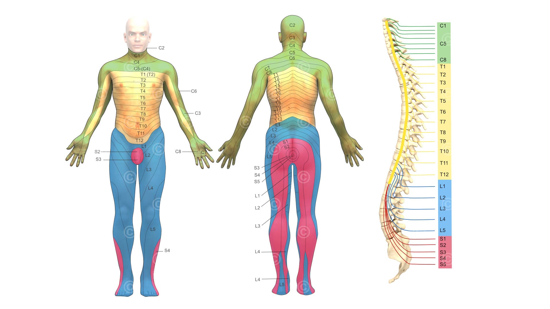
Creation of illustrations and graphics for use in educational materials and training courses in the field of health and fitness training. 3D illustration of the peripheral nervous system with color assignment of the treated areas to the respective exit point of the nerves from the spinal column.
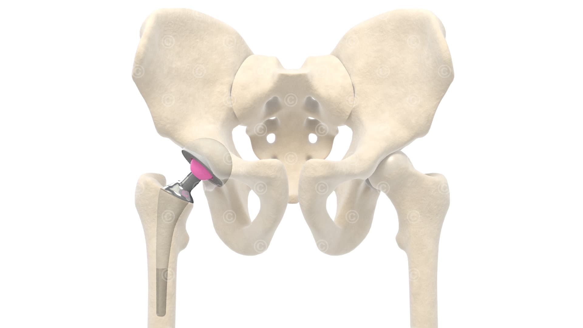
3D illustration of an endoprosthesis of the hip, ie the use of an artificial condyle of the femur and an artificial socket at the hip.
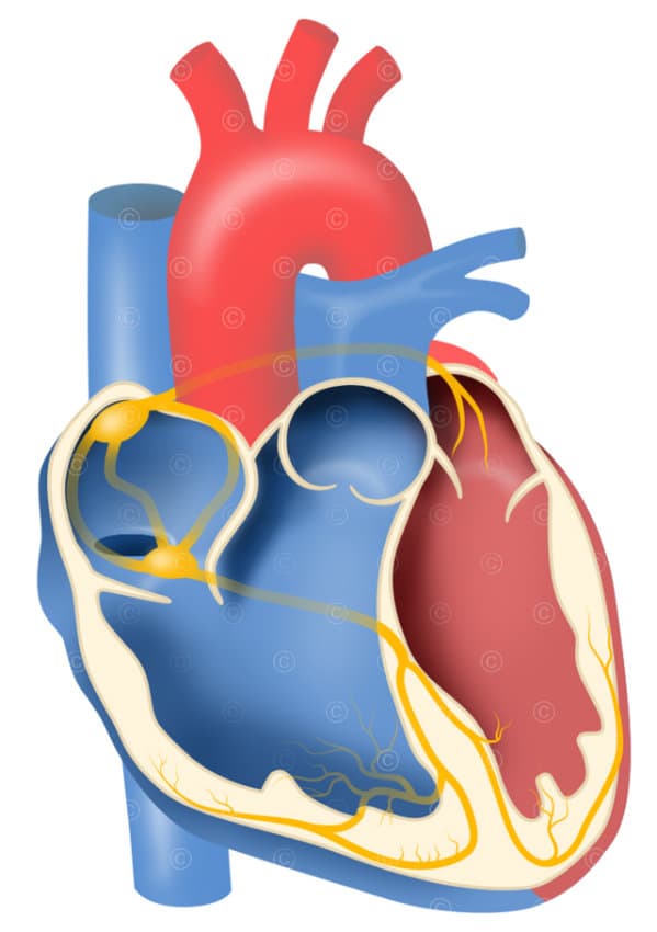
Medical graph of cardiac conduction system: Atrioventricular node (AV node), sinus node, HIS bundle, right and left chamber thigh, Purkinje fibers.
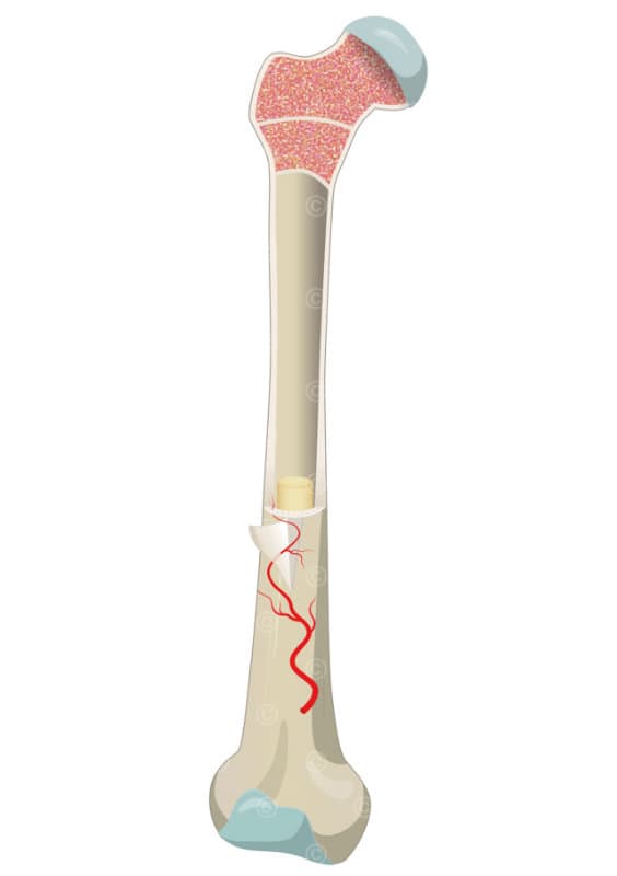
Graphic on the anatomical structure of bones using the example of a long bone (in the example femur): bone tissue, periosteum, marrow cavity, supplying blood vessels, articular surface, red and yellow marrow, epiphyseal plate.
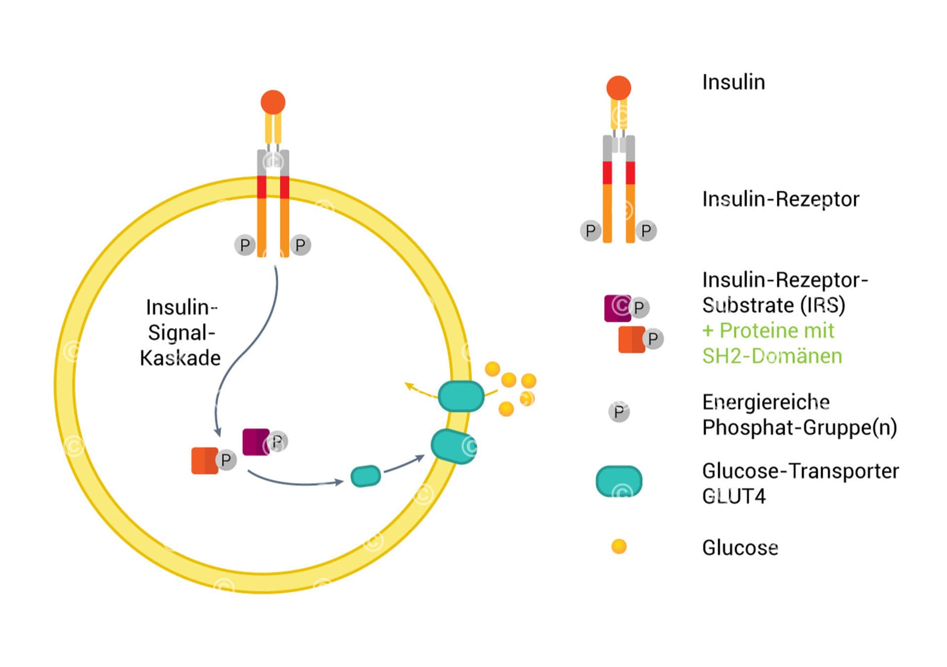
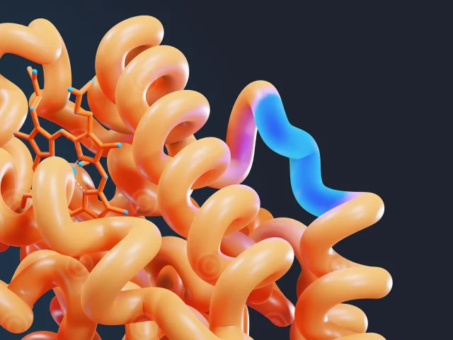
Graphic to a simplified insulin signal cascade.
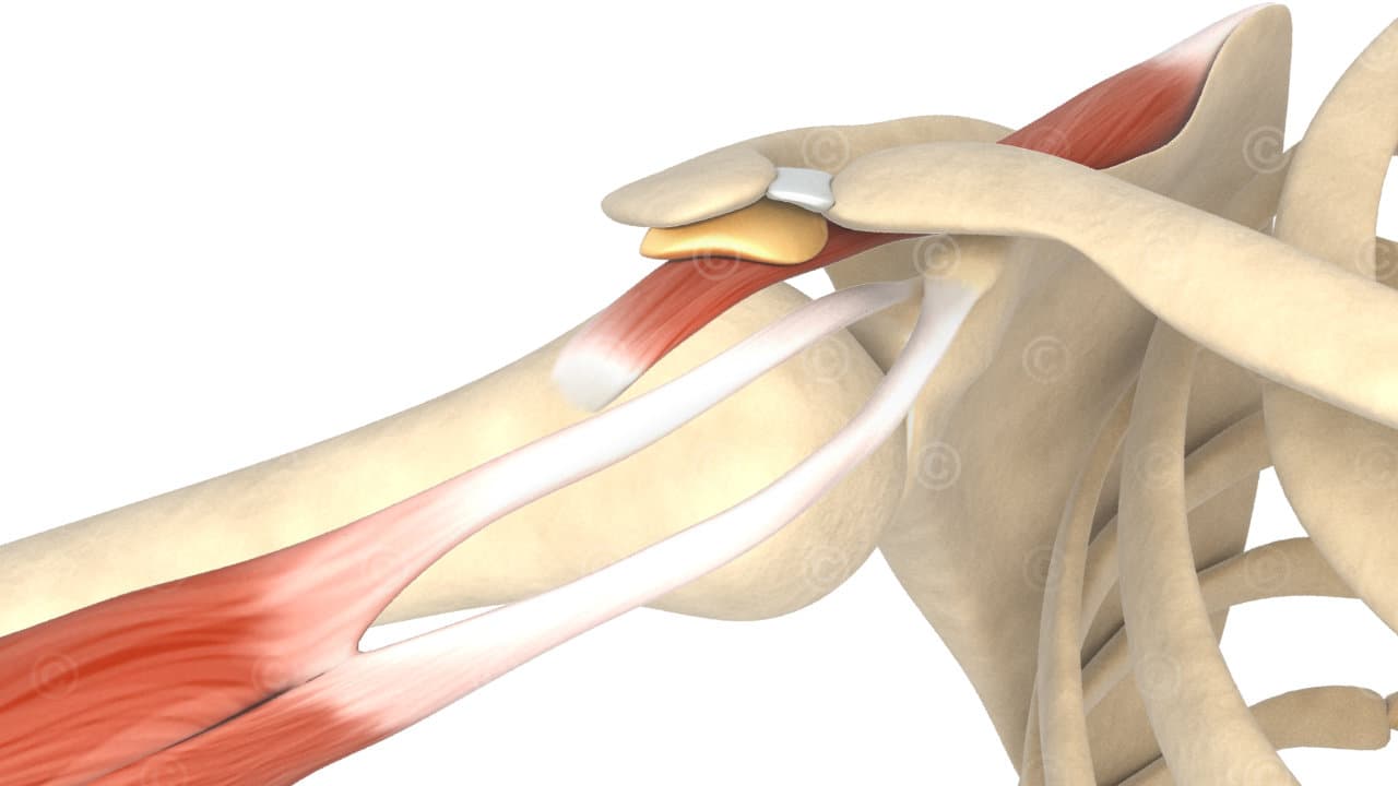
Illustration of the impingement syndrome of the shoulder: pinching the bursa and the supraspinatus tendon between the acromion (bone protrusion of the scapula).
Graphic on the role of dopamine in Parkinson’s disease: The brain uses the neurotransmitter “dopamine” to fine-tune muscle movements. When the available dopamine has dropped by 70 to 80% of normal, Parkinson’s symptoms appear. A regulation of the muscles is no longer possible.
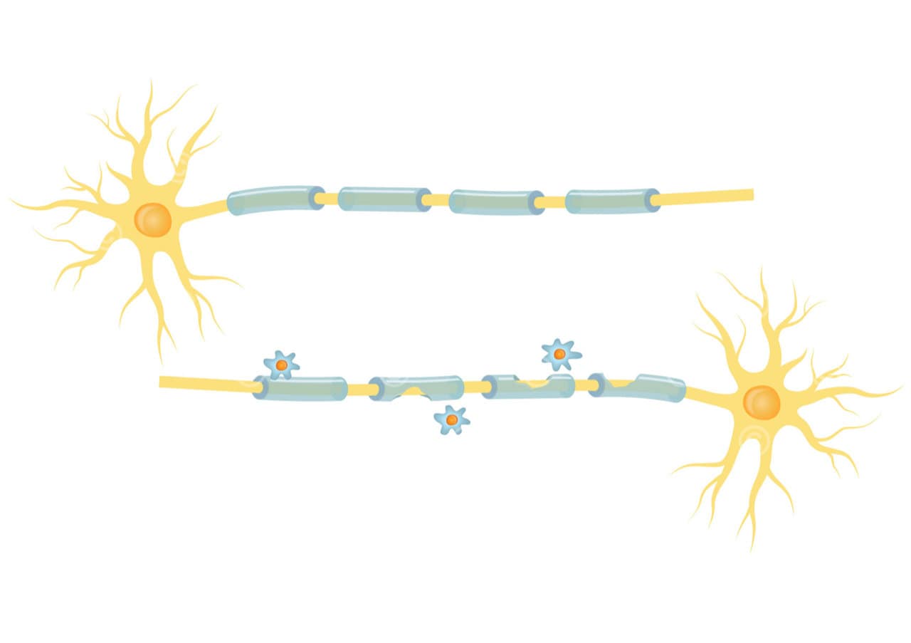
Graphic showing the degradation of the myelin layer of the nerve cells in a multiple sclerosis disease. The damage to the myelin sheath disturbs the transmission of nerve impulses.
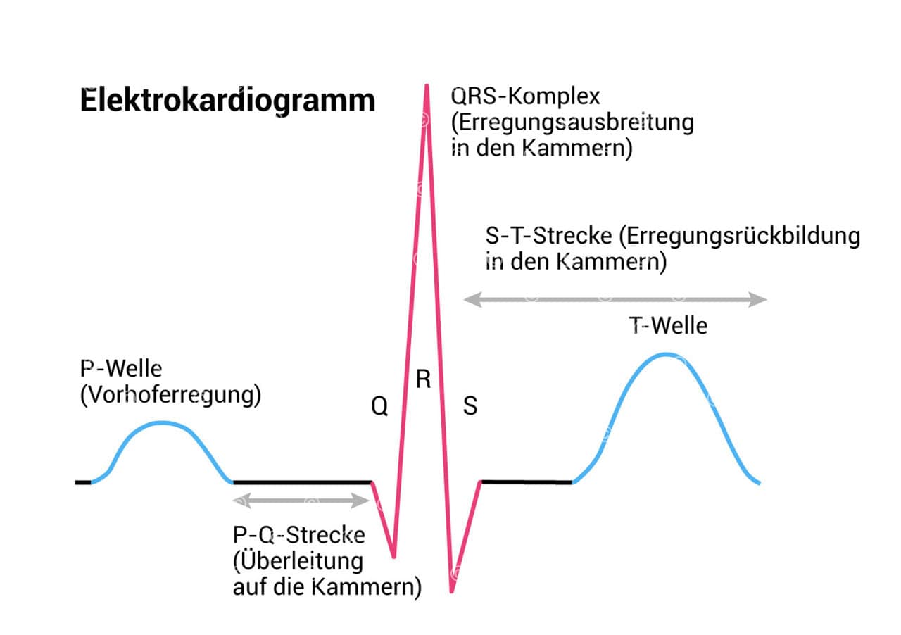
Electrocardiogram (ECG) section graph: P wave, PQ path, PQ duration, QRS complex, J point, ST distance, T wave, QT duration, U wave.
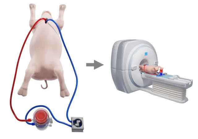
Project description:
Content: 9 illustrations
Utilization: Teaching Materials, Website, PowerPoint
Specifications: DIN A4 (3508*2480px)
Client: IST Studieninstitut
The rights of use of the illustrations shown are with the respective clients. The images are protected with a watermark – use is prohibited.