Anatomy
Illustration suprahyoid muscles
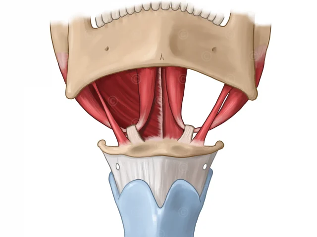
Design of an antomic illustrations of the suprahyoid muscles: M. digastricus, M. mylohyoideus, M. geniohyoideus, M. stylohyoideus.
Design of an antomic illustrations of the suprahyoid muscles: M. digastricus, M. mylohyoideus, M. geniohyoideus, M. stylohyoideus.
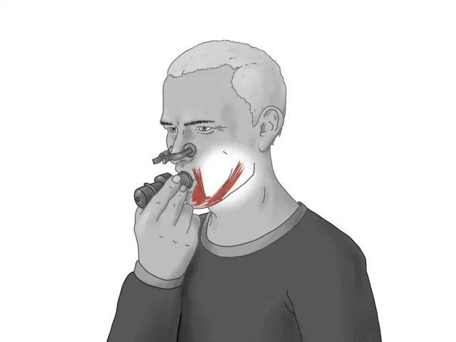
Illustrations of three therapeutic exercise for the treatment of dysphagia: CTAR, Shaker, and EMST.

Various drawn illustrations about different types of tracheostomy management on dysphagia treatment for a medical publication.

Illustrations of catheter-based embolization of a carotid artery fistula (CSCF) by access via the superior ophthalmic vein (SOV).

Illustration of a videofluoroscopic swallowing study – VFSS

Illustrations of the nerves involved in the swallowing process and the muscles they innervate.

Illustrations of a study on the accumulation of lymph node metastases in the vicinity of colorectal tumors.

Design of series of illustrations on dysfunctional swallowing processes in dysphagia in the style of endoscopic swallowing examination (FEES).

Illustrations of reduction of mitral regurgitation by suturing the mitral valve leaflets (Alfieri stitch).
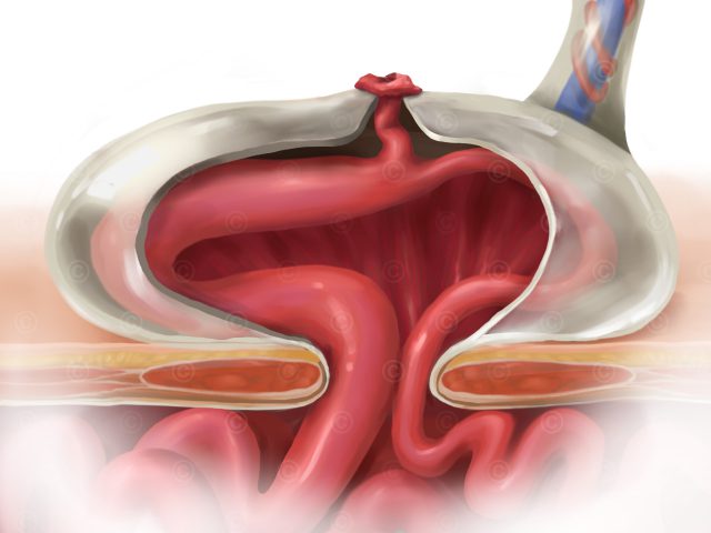
Drawn illustration of omphalocele malformation with omphalomesenteric duct for a medical publication.