Background
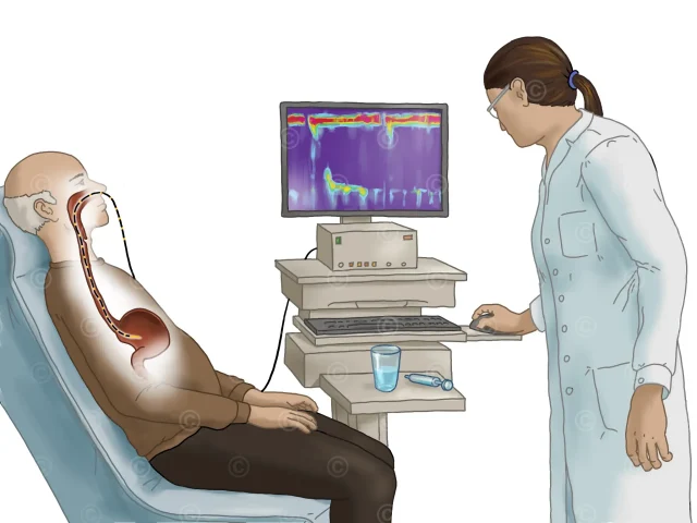
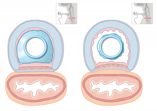
In a videofluoroscopy, the patient’s swallowing process is recorded over time (25-30 images per second) using X-rays. The swallowing samples are mixed with contrast agents to make them visible in the X-ray image and to identify possible causes of swallowing disorders (dysphagia).
Project description
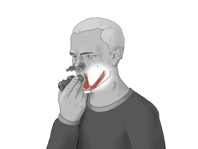
Design of an illustration for performing a videofluoroscopic examination of swallowing (videofluoroscopy, VFSS). The illustration shows a sitting patient with an X-ray machine positioned to the side. The examiner – on the right of the picture – is wearing blue X-ray protective clothing and is administering swallowing samples to the patient with a spoon, which are enriched with contrast medium.
Project details:
Content: 1 illustration
Utilization: Scientific publication by Bendix Labeit, M.D.
Specifications: DIN A5, Landscape
Client: Dr. med. Bendix Labeit – Klinik für Neurologie mit Institut für Translationale Neurologie Universitätsklinikum Münster (UKM)
The rights of use of the images shown here belong to the client, use is not permitted. The images are protected with watermarks.