Background
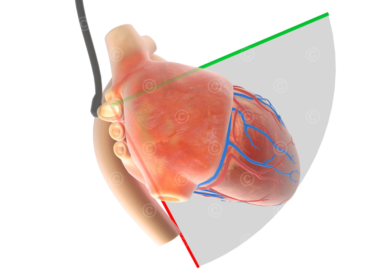
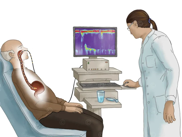
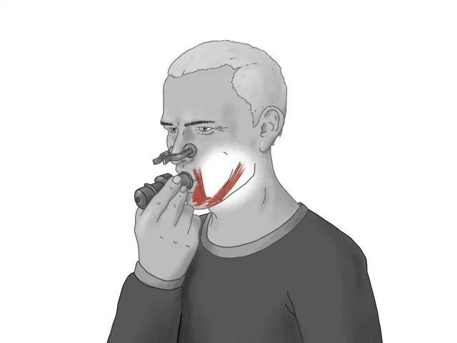
Transoesophageal echocardiography (TEE) is a special form of ultrasound examination of the heart in which the transducer is not placed on the chest wall but in the oesophagus. This technique enables a particularly detailed and precise image of the heart and its structures, as the transducer is positioned relatively close to the heart in direct contact with the esophagus. Depending on the position in the oesophagus and the rotation of the transducer, different structures of the heart can be detected.
Project description
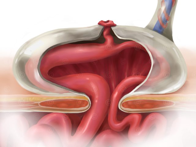
Design of illustrations about the position of the ultrasound transducer and the orientation of the transducer plane in a ME-LAX (midesophageal-long-axis) view. This standard view in transesophageal echocardiography provides a view of the left atrium, left ventricle, mitral valve, left ventricular outflow tract, aortic valve, and ascending aorta.
Two images were created with the heart closed and the sound plane transparent, illustrating how the transducer must be positioned and oriented in order to achieve correct imaging with the ultrasound view. In the other images, the lower part of the heart was resected to allow correlation with real ultrasound images from a midesophageal-long-axis view.
Project details:
Content: 4 illustrations
Utilization: Training courses of the association
Specifications: DIN A5, transparent background
Client: Dr. Dieter Wally, Verein zur Förderung der perioperativen Notfallechokardiographie
The rights of use of the images shown here belong to the client, use is not permitted. The images are protected with watermarks
Illustrations