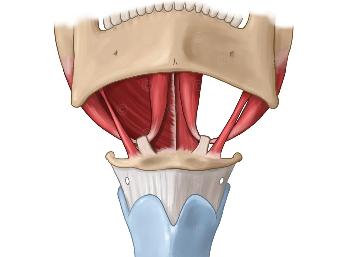
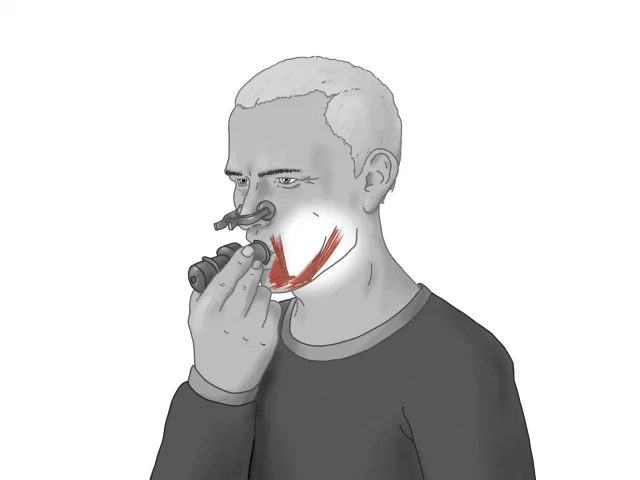
Design of an antomic illustrations of the suprahyoid muscles. These muscles are a group of four muscles in the throat area located above the larynx. They play an important role in the movement of the larynx and hyoid bone, as well as in supporting the swallowing function.
The four suprahyoid muscles are:
Musculus digastricus: This muscle runs from the mandible to the hyoid bone and is responsible for elevating the hyoid bone and larynx.
Stylohyoid muscle: This muscle runs from the temporal bone to the hyoid bone and assists the digastric muscle anteriorly and posteriorly in lifting the hyoid bone.
Musculus mylohyoideus: This muscle runs from the mandible to the hyoid bone and is responsible for lifting the hyoid bone. It also assists the digastricus anterior and posterior muscles in lifting the hyoid bone. On the (patient) left side of the illustration, the mylohyoid muscle has been removed to reveal the geniohyoid muscle behind it.
Musculus geniohyoideus: This muscle is a slender, wedge-shaped muscle in the throat area located between the mandible and the hyoid bone. It is part of the suprahyoid musculature and plays an important role in moving the hyoid bone and hyoid bone, as well as supporting swallowing function.
Together, these muscles work to move the larynx and hyoid bone and support the act of swallowing. Weakness or paralysis of the suprahyoid muscles can lead to impaired swallowing function, which is called dysphagia.
Project details “Illustration suprahyoid muscles”:
Content: 1 Illustration
Usage: Medical reference book
Technisches: DIN A5
Client: Dr. Dziewas, Dr. Warnecke, Universitätsklinikum Münster
The rights of use of the images shown here belong to the client, use is not permitted. The images are protected with watermarks
Illustration suprahyoid muscles: