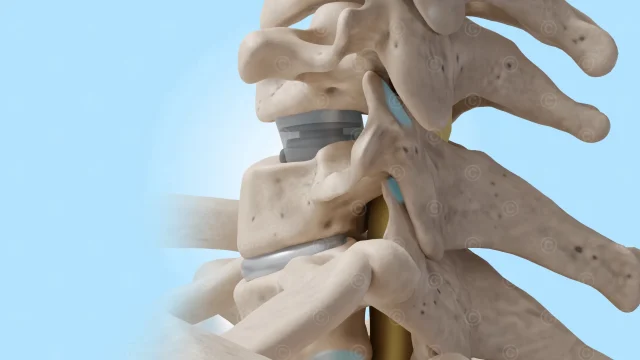
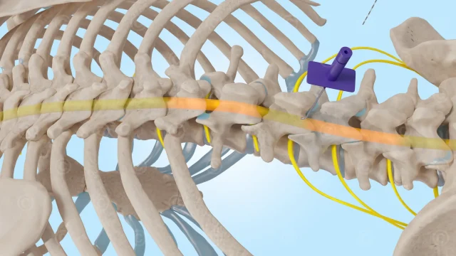
Background
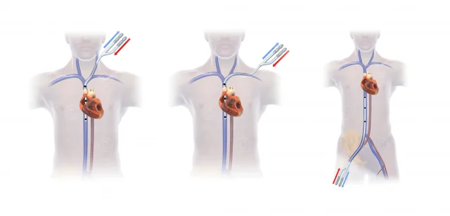
A carotid fistula (CSCF – carotid-cavernous sinus fistula) is an unintentional connection between the carotid arteries and the cavernous sinus. These fistulas are usually caused by trauma such as skull base fractures or vascular disease. As a result, blood flows from the artery directly into the venous system. As a result, the blood pressure in the veins rises and normal blood circulation is disrupted, leading to various symptoms.
Project Description
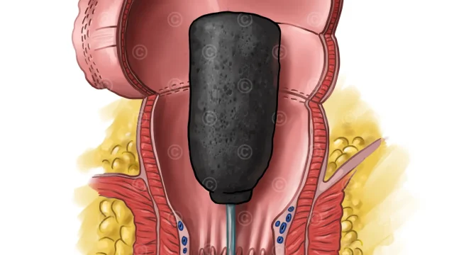
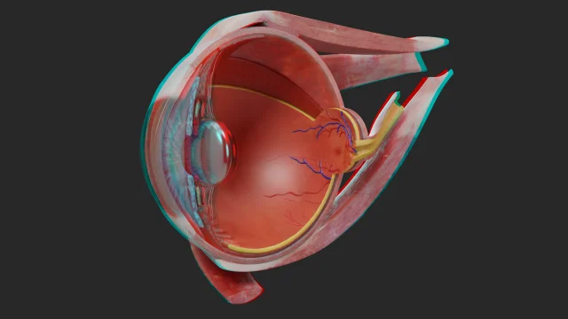
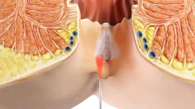
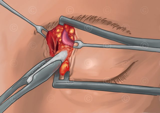
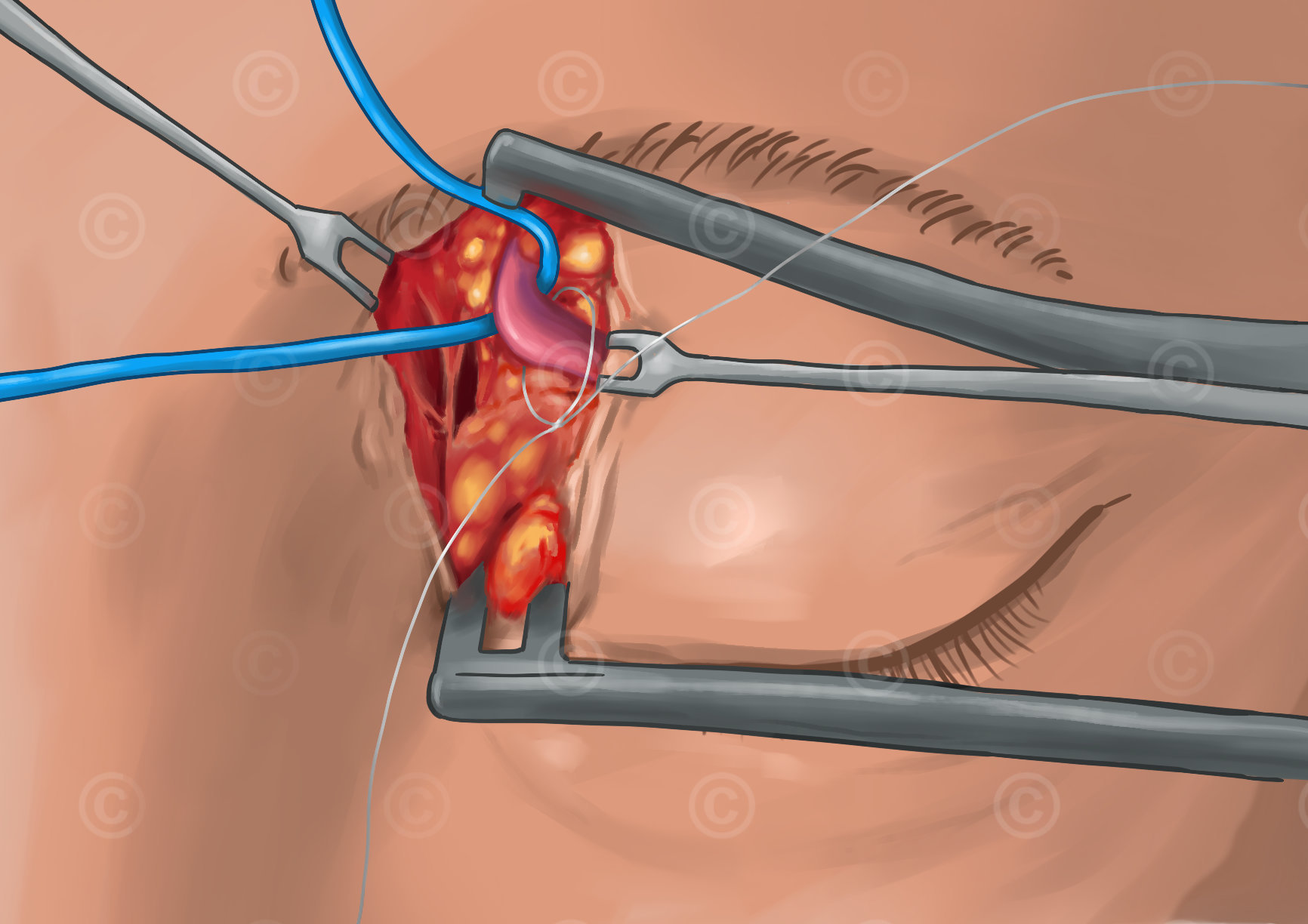
For the medical textbook “Endoscopic Transorbital Surgery of the Orbit, Skull Base and Brain” we designed illustrations to explain the surgical procedures for catheterizing a carotid fistula. To treat the fistula, a catheter is inserted through the superior ophthalmic vein (SOV) and advanced to the site in the brain where the fistula is located. An incision is made medial/cranial to the eye and the superior ophthalmic vein is exposed and prepared. A catheter is inserted into the vein and advanced to the fistula in the cavernous sinus.
An additional illustration shows the anatomy of the eye with the surrounding veins and two possible approaches to the fistula via the superior ophthalmic vein (SOV approach) or the inferior ophthalmic vein (IOV approach).
Project details:
Content: 5 drawn illustrations
Utilization: Book “Endoscopic Transorbital Surgery of the Orbit, Skull Base and Brain” – Publisher: Springer Nature – Editors: Theodore H. Schwartz, Doo-Sik Kong, Kris S. Moe.
Specifications: Resolution 2K and 4k
Client: Prof. Dr. med.Maximilian Linxweiler / Universität des Saarlandes / Springer Nature
The rights of use of the images shown here belong to the client, use is not permitted. The images are protected with watermarks.
Illustrations


Screencast drawing anatomy ophthalmic veins:
The rights of use of the image material lie with the client.