Project description:
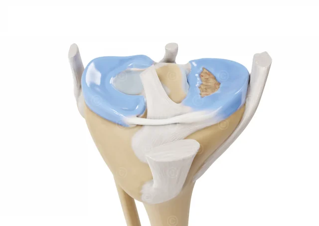
Design of three anatomical illustrations of the knee joint for a manufacturer of lasers and laser fibers (Biolitec). Since the illustrations were to be used in a wide variety of applications, images were created in very high resolution (DIN A1 – 300dpi).
The first illustration visualizes pain experienced by patients with a knee replacement in a slightly lateral view.

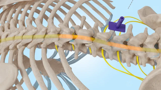
The second illustration shows the anterior nerves of the knee joint (superolateral genicular nerve, superomedial genicular nerve, inferolateral genicular nerve, inferomedial genicular nerve, recurrent fibular nerve, and nerves to the vastus lateralis and medialis).

The third illustration shows the knee joint with the fibula (calf bone), tibia (shin bone), and femur (thigh bone) in a transparent leg.

Project details illustration knee replacement and nerves knee
Content: 3 illustrations
Use: Brochures, trade fair booth, website, print, social media
Specs: DIN A1
Client: Biolitec
The rights of use for the images shown here belong to the client; use of the images shown here is not permitted. The images are protected by watermarks.