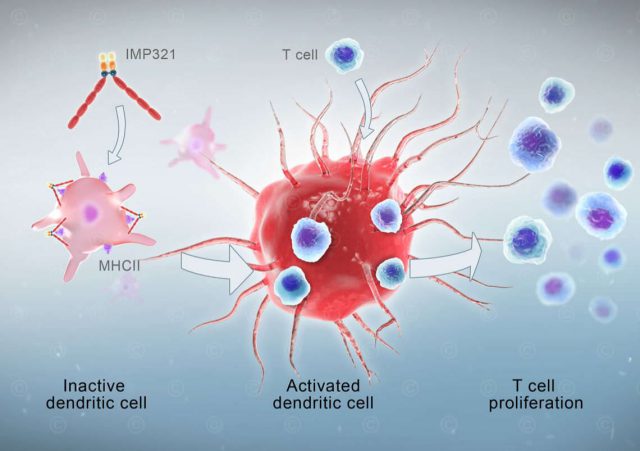
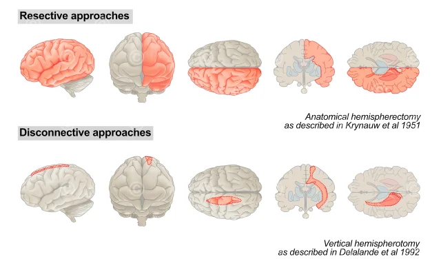
Illustrations proteins

Licensable illustrations of various proteins such as hemoglobin, antigen, SARS spike glycoprotein, interferon, etc.

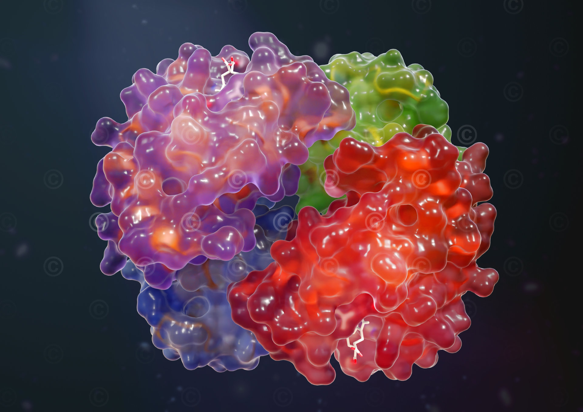
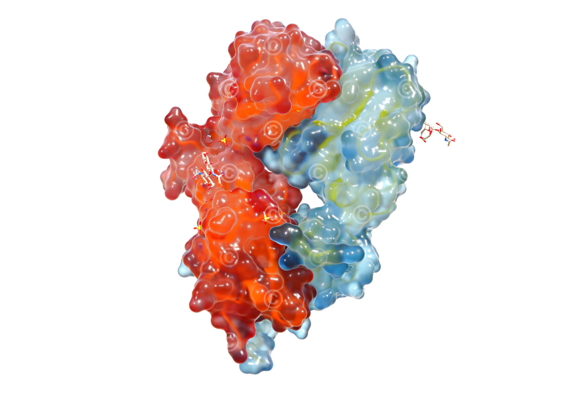
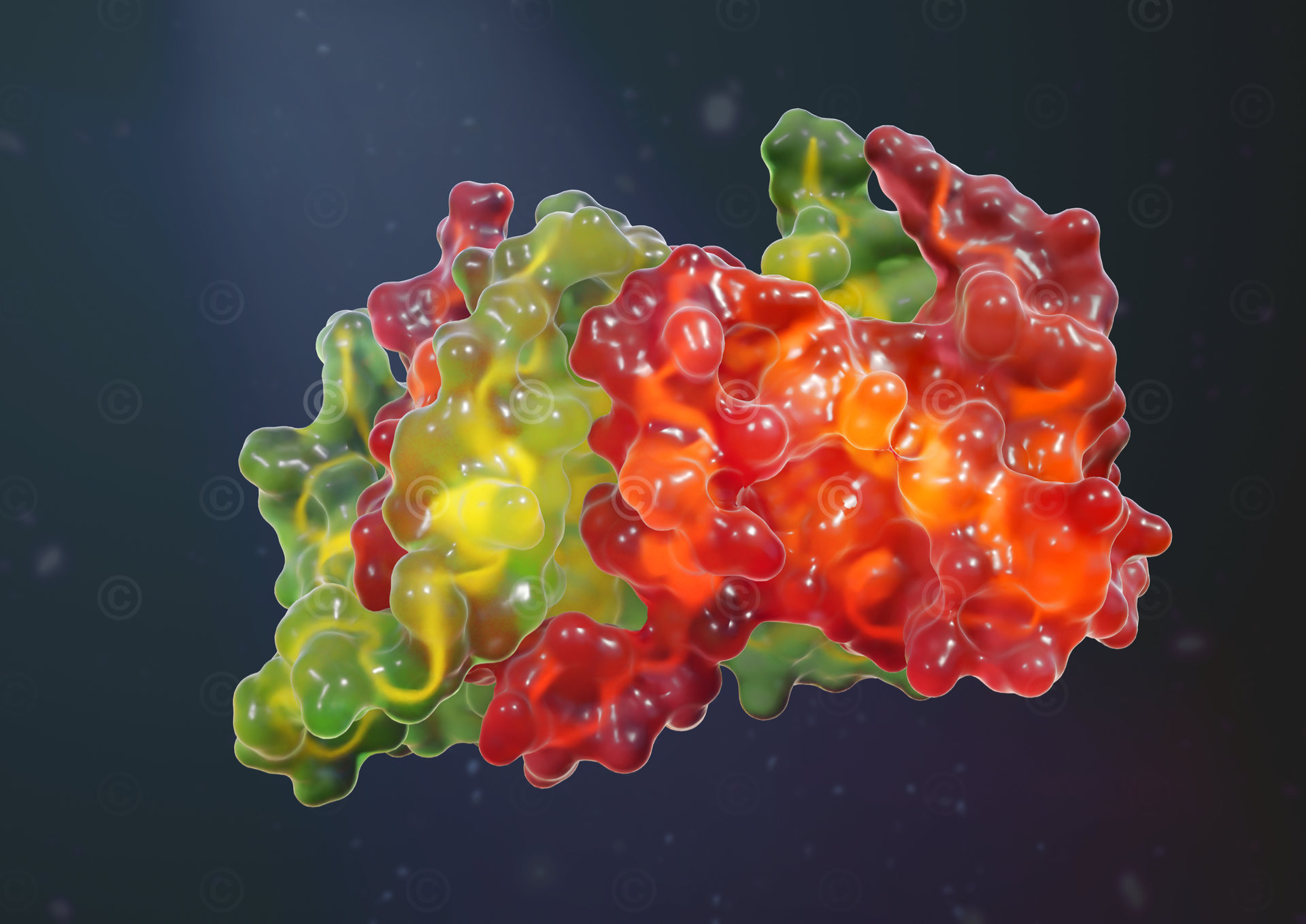
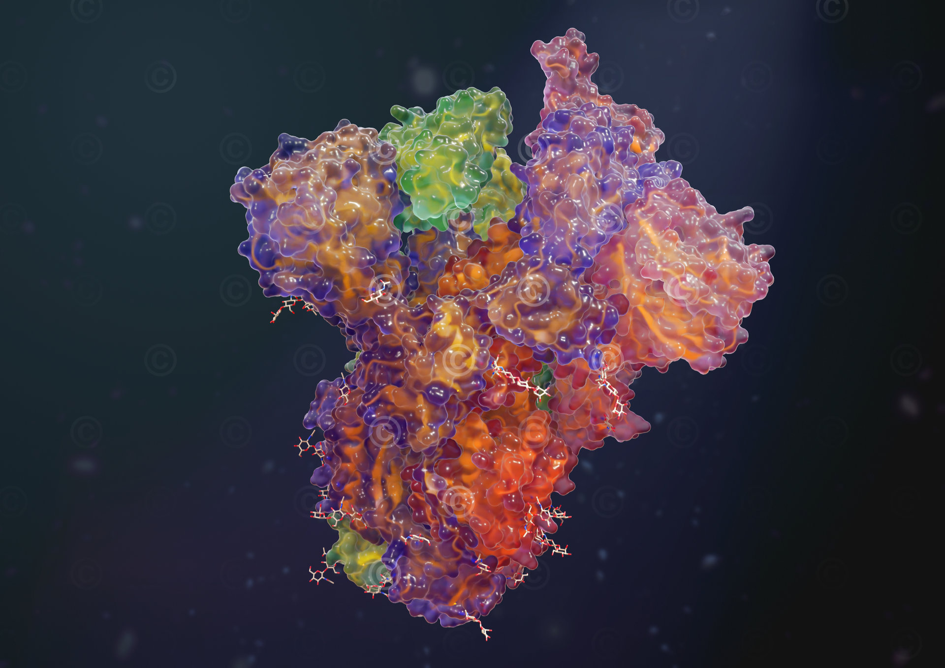
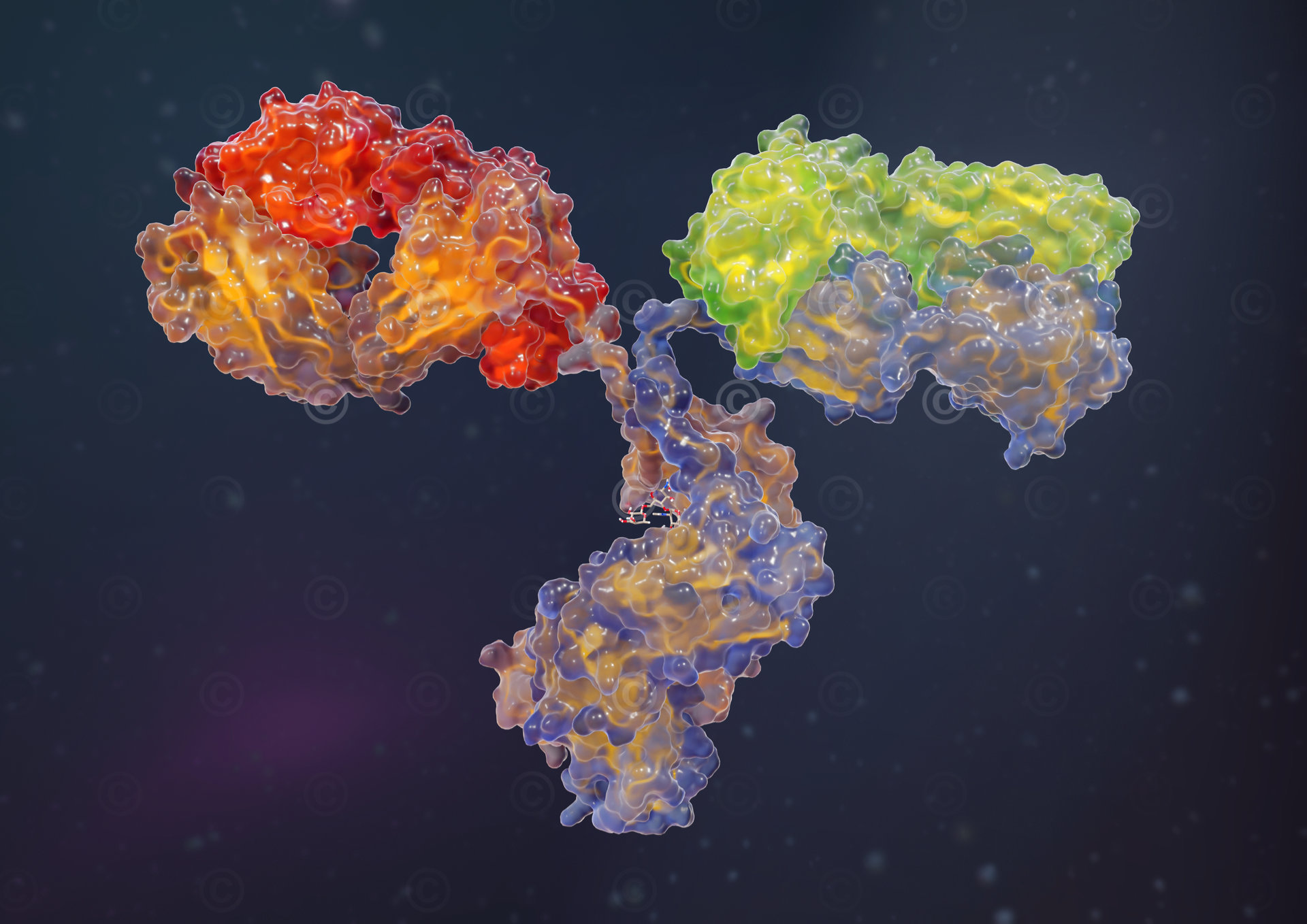
Design of illustrations of different proteins based on structural data of the “Protein Databank”. The underlying idea was to get ahead of the conventional rather technical representation (surface model, ribbon model, etc.) of the proteins and to create appealing and impressive illustrations of the structures that have a high recognition value. When developing the “look” of the illustrations, we had a candy look in mind, i.e., a colorful design that is reminiscent of candy and has a positive connotation. The illustrations were designed in two versions: the first version shows the protein on a dark background that indicates the intracellular environment and emphasizes the colors and structures. The second version shows the protein on a neutral white background for optimal integration, for example, in the context of a printed text.

You will find these illustrations in a smaller version with our logo in our category “Free Illustration“. These are free to use (even commercially) under a a Creative Commons license. Please note the terms of use described there. If you are interested in purchasing the images in higher resolution and/or with further rights of use, visit our Gumroad-Shop (external link).

Project details: 

Content: 21 illustrations

Utilization: free illustrations, Gumroad, Stock Photo

Specifications: DIN A3 / 300dpi (4961×3508 pixel)

Client: MedicalGraphics

The rights of use for the illustrations shown here lie with the client; use is not permitted. The images are protected with watermarks. If you are interested in the illustrations, please use the versions of the images in the rubric “Free Illustrations” under the terms of use mentioned there or acquire a license via Gumroad.
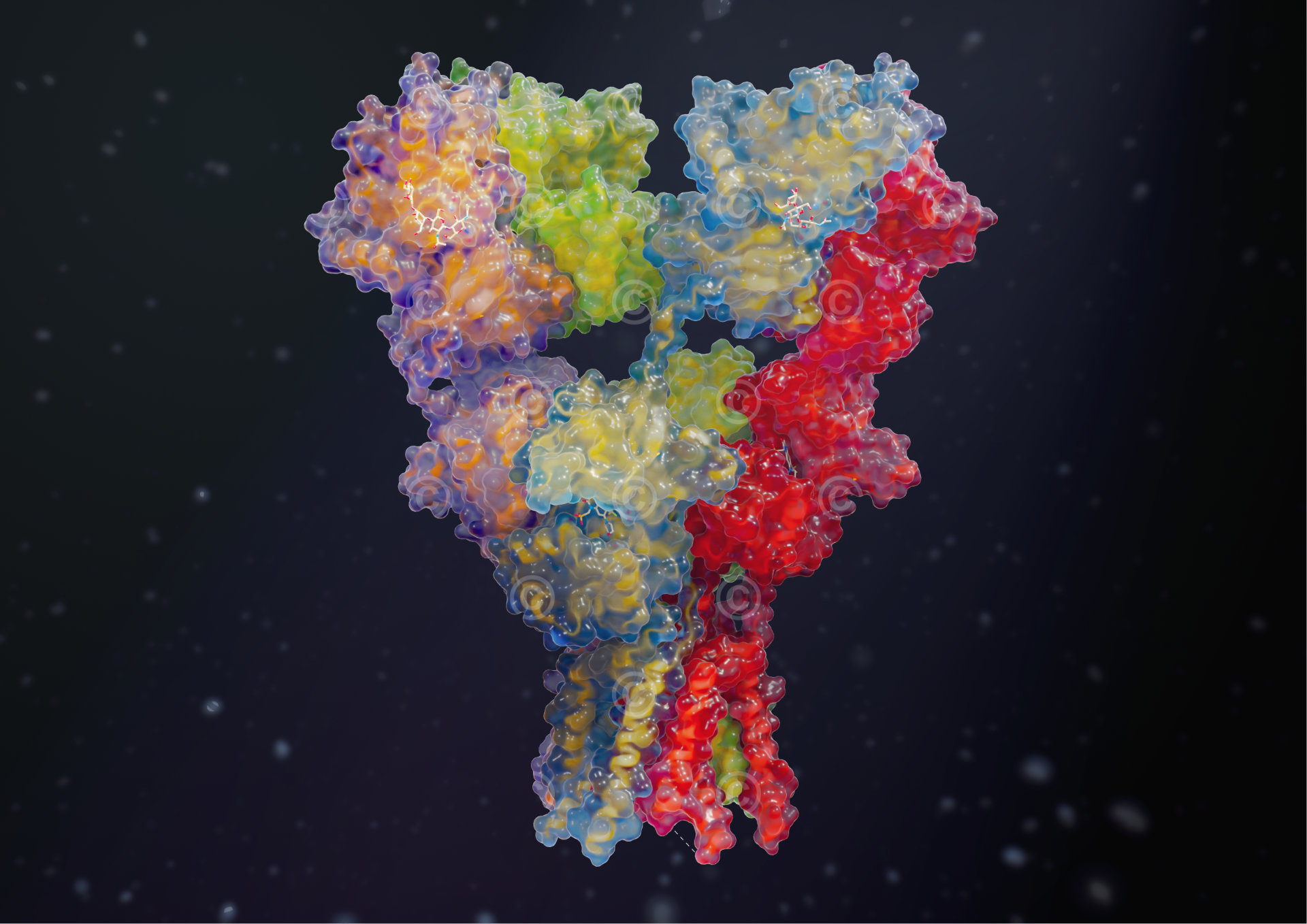
ampa receptor dark
AMPA Receptor

AMPA receptors (a-amino-3-hydroxy-5-methyl-4-isoxazolepropionic acid receptor) belong to the glutamate receptors and are the most common neurotransmitter receptors in the central nervous system. The upper part of the receptor is located extracellularly and recognizes and binds glutamate. The lower part is located in the membrane and, after binding glutamate, forms a channel through which ions pass through the membrane. – This illustration is based on structural data from Sobolevsky, A.I., Rosconi, M.P., Gouaux, E.

hemoglobin dark background
Hemoglobin

Hemoglobin is the protein complex of red blood cells (erythrocytes) whose task is to bind oxygen and carbon dioxide and thus transport them in the bloodstream. – The illustration is based on structural data from Perutz, M.F.

t zell receptor
T cell receptor

T cell receptors are located on the surface of T cells and are responsible for recognizing antigens presented by MHC molecules. It can recognize foreign antigens and in this case activate the T cell. – The illustration is based on structural data from Garcia, K.C., Degano, M., Stanfield, R.L., Wilson, I.A.

interferon beta dunkler hintergrund
Interferon Beta

Beta-interferon is produced by virus-infected fibroblasts in response to the recognition of viral or bacterial nucleic acid. It activates surrounding cells in which proteins are produced that inhibit further (viral) protein synthesis in those cells and promote the degradation of viral and cellular RNA. – The illustration is based on structural data from Karpusas, M., Nolte, M., Benton, C.B., Meier, W., Lipscomb, W.N., Goelz, S.

sars cov2 spike protein
SARS-COV-2 Spike Protein

The SARS spike glycoprotein spreads over the surface of the SARS-CoV-2 virus and docks onto the ACE2 receptors of the cell surfaces of the lung tissue to infect them. – The illustration is based on structural data from Kirchdoerfer, R.N., Wang, N., Pallesen, J., Turner, H.L., Cottrell, C.A., McLellan, J.S., Ward, A.B.

antigen
Antigen

More projects: