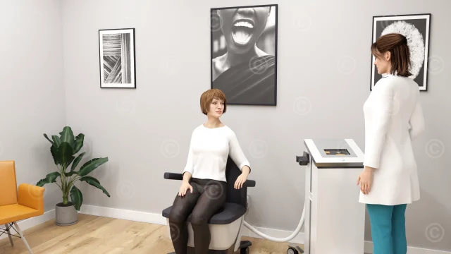
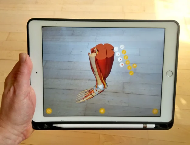
Creation of graphical content for a web-based training of Allianz AG on the musculoskeletal system of the upper body with a focus on the elbow joint. A linear, seamless animation leads through the topics of the e-learning. At defined points, the animation stops and textual information, illustrations and interactive 3D models are displayed to explain the content, or the viewer’s level of knowledge is tested in a quiz.
This e-Learning module provides basic knowledge of elbow anatomy and common disorders and injuries.
Rights of use of the visual material: Allianz AG
Music: Kevin McLeod – Impact prelude – License: Creative Commons (CC BY 3.0)
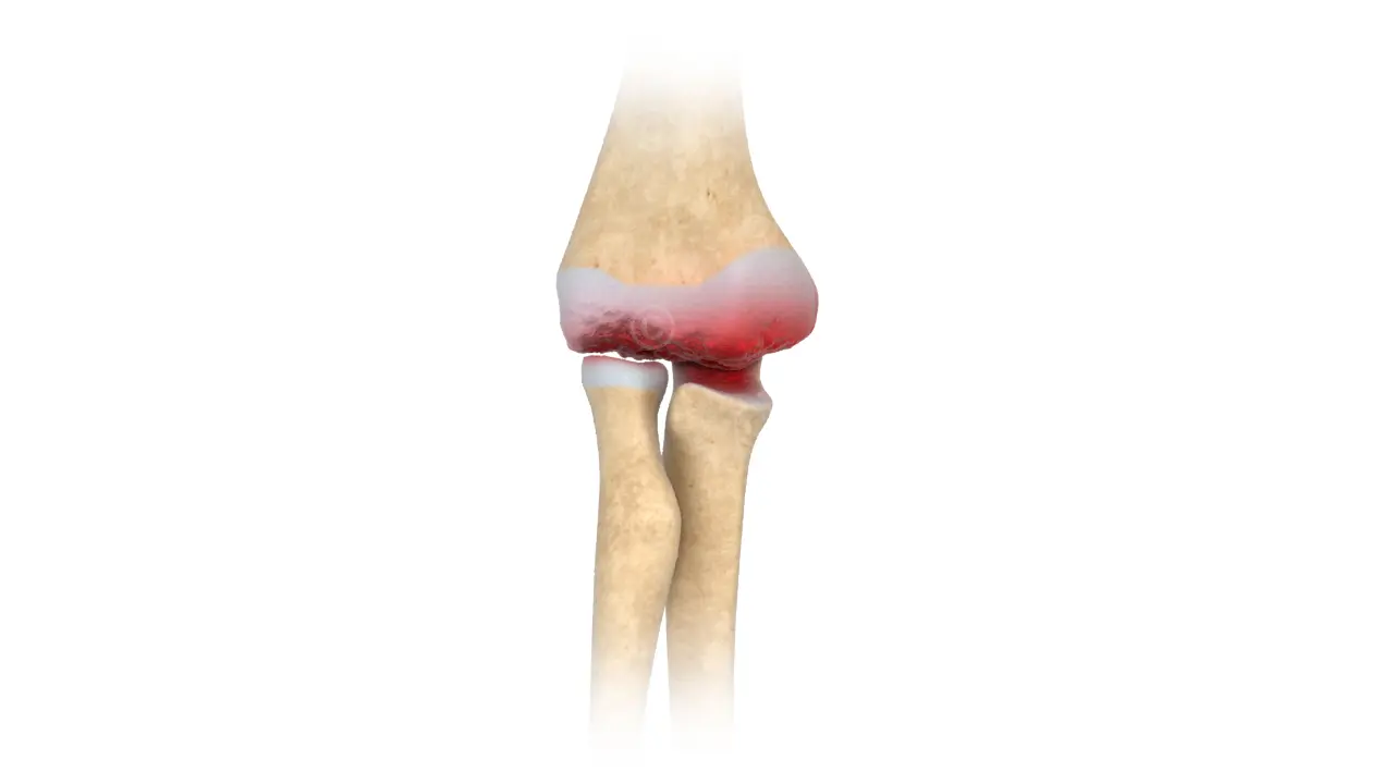
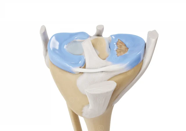
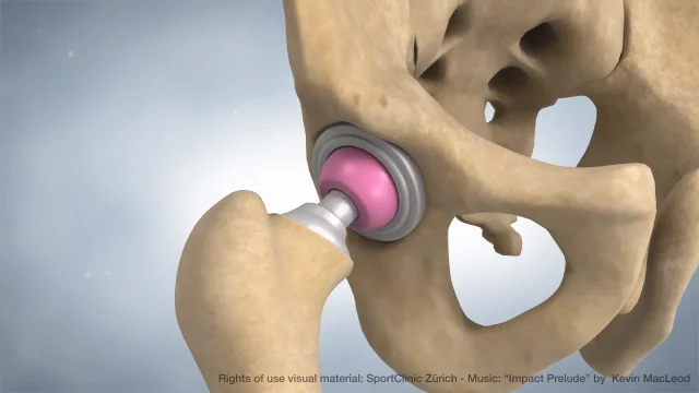
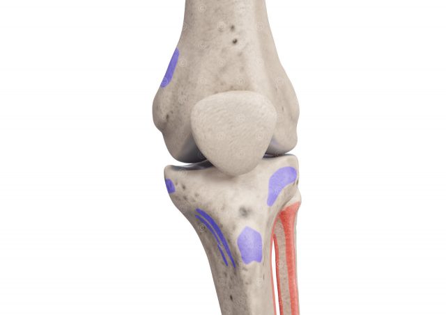
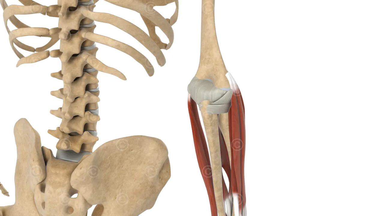
The elbow is a joint that connects the humerus, the ulna and radius and consists of the following anatomical structures: humerus, radius, ulna, joint capsule, joint ligaments, joint bursa (bursae), muscles (biceps, triceps, brachialis and brachioradialis etc.). The elbow is an important part of the arm that allows flexion (bending) and extension (stretching) of the forearm. It plays a crucial role in performing everyday movements such as grasping, lifting and moving objects. The complex anatomy of the elbow contributes to the stability and functionality of this joint.
The following disorders of the elbow were addressed in the e-Learning:
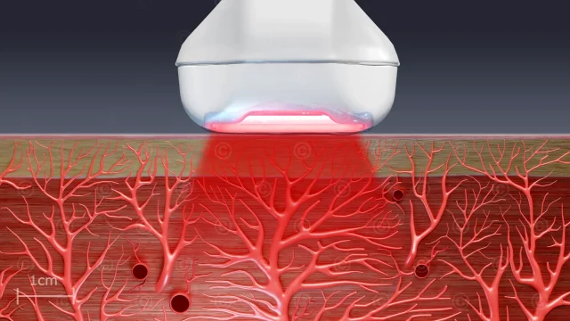
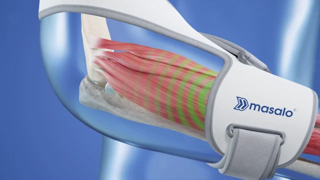
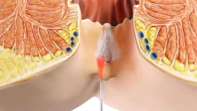
Epicondylitis, also known as or tennis elbow, is a painful condition that results from overuse or repetitive strain on the tendon insertions in the elbow. Epicondylitis most commonly occurs on the outer side of the elbow, where the wrist and finger extensor tendons are located on the lateral epicondyle of the humerus. Symptoms of epicondylitis include pain and tenderness at the outer or inner elbow that may worsen when reaching, lifting or twisting the forearm and hand. Sometimes the pain radiates into the forearm. Epicondylitis is most often treated with a combination of conservative measures to relieve the pain and promote healing.
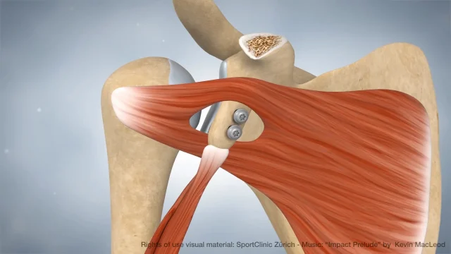
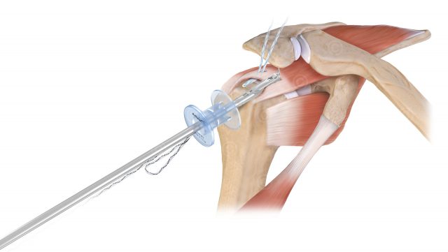
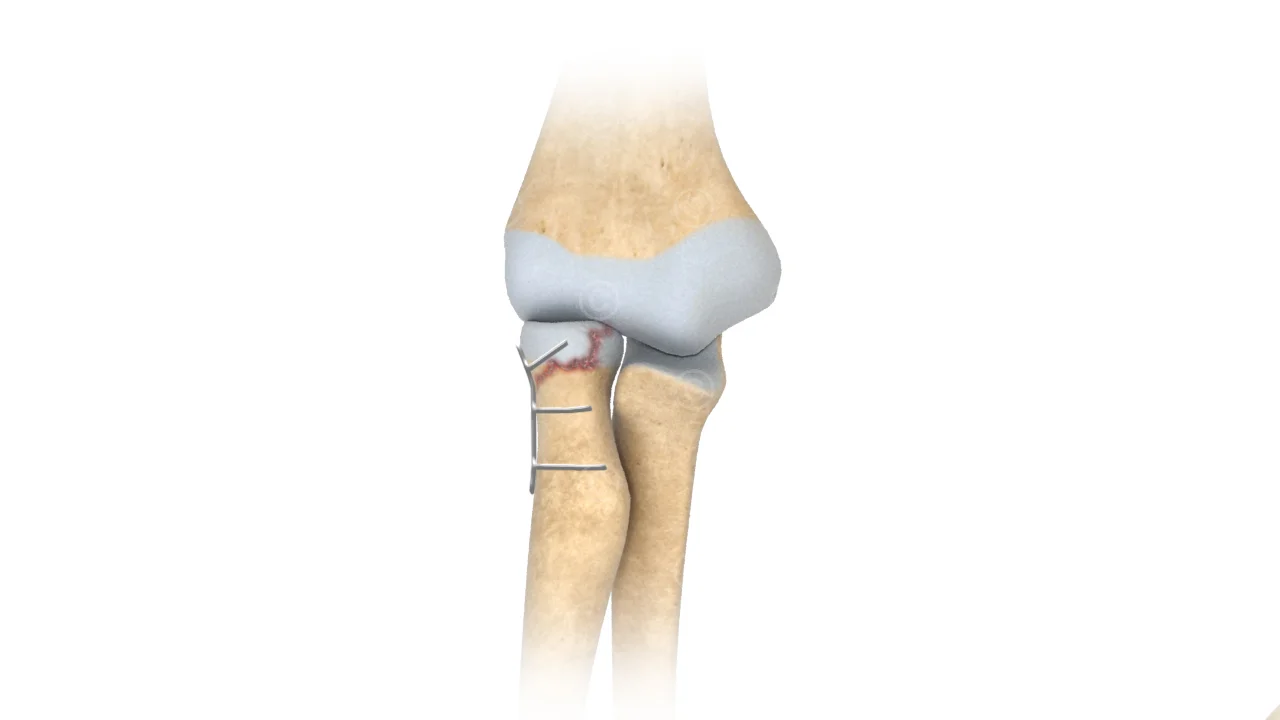
Fractures in the elbow are often caused by falls on the elbow joint or by a fall on the outstretched arm. The fractures are often so-called comminuted fractures, for the treatment of which surgical intervention is usually necessary. Here, the individual bone fracture pieces are brought into their original position and then fixed with wires or by using a plate.
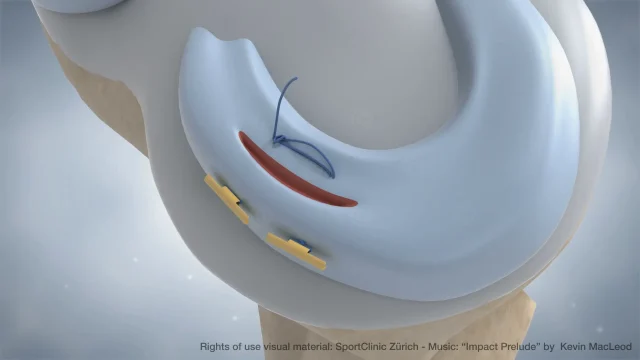
Rarely, patients develop Osteoarthritis of the Elbow. In its course, the articular cartilage degrades until the elbow joint surfaces rub against each other painfully. In addition, joint mice can form. These are small cartilage or bone fragments that further restrict the normal movement of the joint.

Project details “E-Learning disorders elbow”
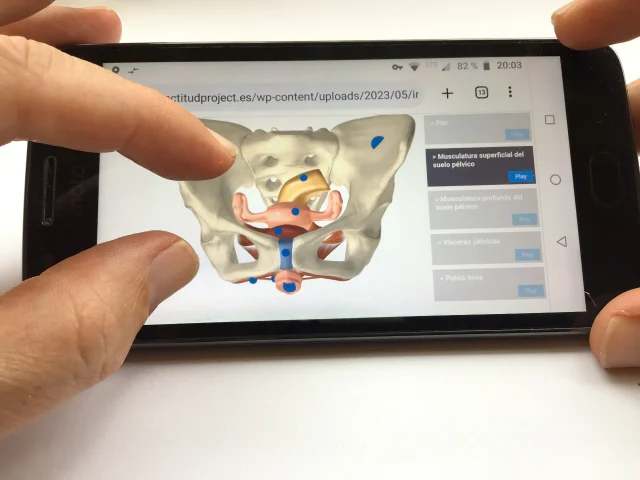
Content: approx. 10 animation sequences, length approx. 25 minutes, approx. 4 illustrations, 1 interactive webGL applications
Use: E-Learning Allianz AG
Specification: Resolution 1280*720px (HD)
Client: Allianz AG / TTS
The rights of use of the visual contents and illustrations shown belong to the customer and are protected by watermarks.
Screencast 3D Modell anatomy elbow:
Rights of use of the visual material: Allianz AG
Music: Kevin McLeod – Impact prelude – License: Creative Commons (CC BY 3.0)
Illustrationen e-Learning elbow