Two projects of Medical Graphics used 3D PDFs to discuss the anatomical structures of 3D models in dialogue with the client. This method has the advantage that the models can be defined more precisely as this would be possible e.g. with pictures. The possibility to explore the 3D models as part of his lectures and trainings convinced the client so much, that the 3D PDFs eventually became a part of the final project.
TEE ultrasound heart:
In the first project illustrations of standard TEE ultrasound levels for the physicians of the “Rober Bosch Hospital” in Stuttgart were created. In a transesophageal echocardiography an ultrasound probe is inserted into the gullet (esophagus), which runs behind the heart and the outgoing vessels. Depending on the position of the probe within the esophagus and its rotation angle, different parts of the cardiac anatomy can be visualized by ultrasound, without disturbing structures such as ribs.
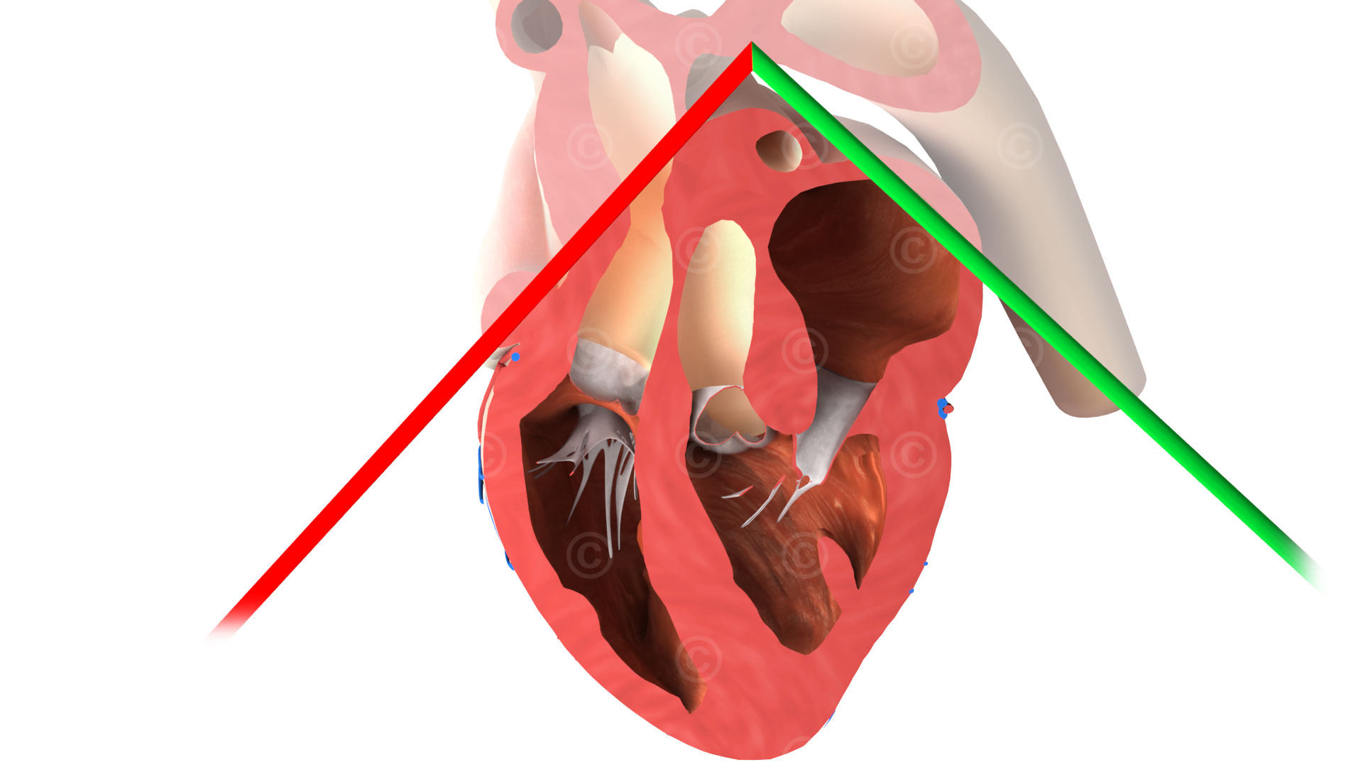
Illustrations of 28 standard section planes were created, which were then used in presentations and trainings. This picutres illustrate how the prope is set to capture a certain part of the heart. Together with the attending physicians in the Rober Bosch Hospital, the respective sectional planes were discussed and then visualized.
The specific orientation of the ultrasound planes presented the greatest challenge. For this purpose, 3D PDFs of a heart with ultrasound planes were created. This worked so well that the doctors decided to use them together with the created illustrations as part of the training.
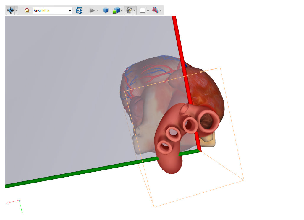
 3D PDF musculoskeletal animation
Animations musculoskeletal system for e-learning:
In the second project 3D PDFs were also actually only used to discuss the anatomical correctness of the 3D models. Here 3D animations should explain the structures of the musculoskeletal system. Again, the customer found the opportunity of 3D PDFs so appealing that he used them in his training and lectures.
In order to view 3D PDFs, a local installation of Acrobat Reader is necessary. Furthermore, most internet browser start a reduced PDF Reader without 3D functionality when selecting a PDF file on the internet. These restrictions were not an obstacle to the above projects. But this was the reason to engaged a little deeper with the possibilities of displaying 3D content on the Internet.
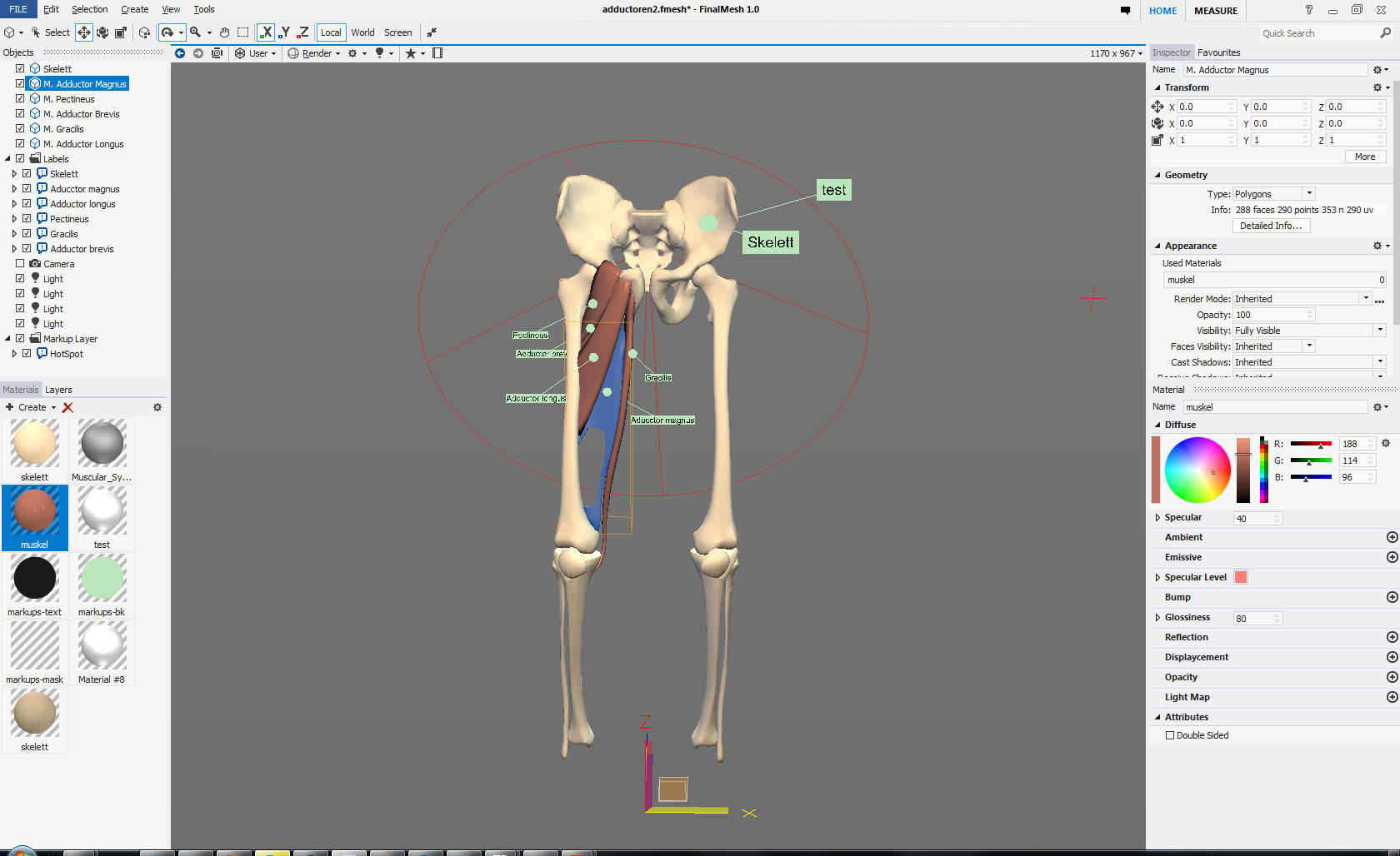
To further evaluate the possibility of real-time 3D content for knowledge transfer, MedicalGraphics test a tool that makes use of the JavaScript API Tree.js for generating WebGL applications. WebGL has the advantage that 3D content can be shown natively (no plugin) in all recent browsers shown. Seamless integration into a website was in the focus. This means: embedding the 3D object directly within a web page and avoiding larger load times. In comparison: the WebGL application below integrates well in a normal web page with a size of 230kb, while our project “WebGL with Unity3D” has a file size of ~40mb (the 3D model is also textured and more detailed).
As an example, an application was created in which a 3D model of the adductors of the right leg were visualized (see below). The explorability of the application makes the course of the muscles visible and its insertion and origin easily understandable.
WebGL App thigh muscles:
Navigation: left mouse button rotation of the model, right mouse button for zoom, middle button for panning the view. More interaction with the 3D model available via the menu at the top left.
The rights of use of illustrations and animations shown lie with the customer.


