Background:
Neurological damage (e.g., ischemic stroke, cerebral hemorrhage) is among the most serious complications in pediatric cardiac surgery. In children with congenital heart defects, both cardiac anatomy and immature cerebral autoregulation of blood flow affect cerebral perfusion.
Project description:
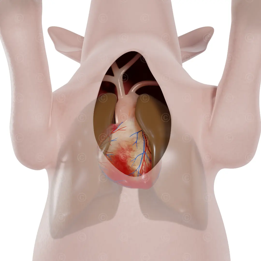
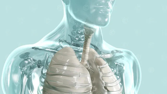
The study “Establishment of an MR-Conditional Porcine Model for Real-Time Assessment of Cerebral Blood Flow During Extracorporeal Circulation” investigates a technique that enables MRI imaging during extracorporeal circulation. Conventional monitoring devices measure only indirect parameters such as blood pressure or blood oxygen saturation. MRI directly reveals any changes in brain tissue. The study focused on its application in pediatric cardiac surgery, as the risk of neurological damage during surgery on a heart-lung machine (HLM) is many times higher in children than in adults.
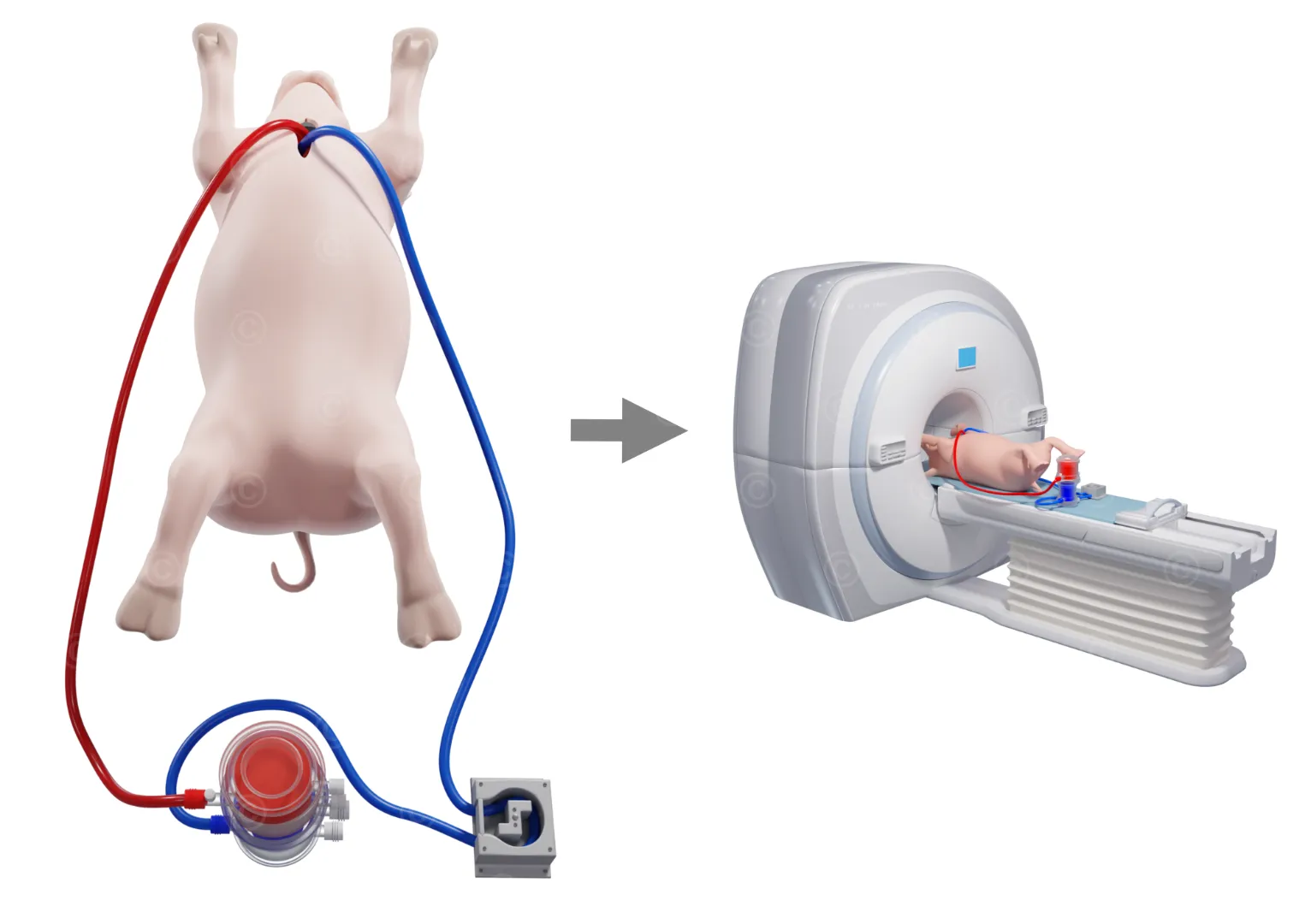
The illustrations showed the layout and configuration of the MRI scanner and the pump unit: while the patient’s head is positioned supine inside the MRI scanner, the roller pump and the oxygenator remains outside the MRI scanner. For this study a “MR-conditional” heart-lung machine was used – that is, a device free of ferromagnetic metals.
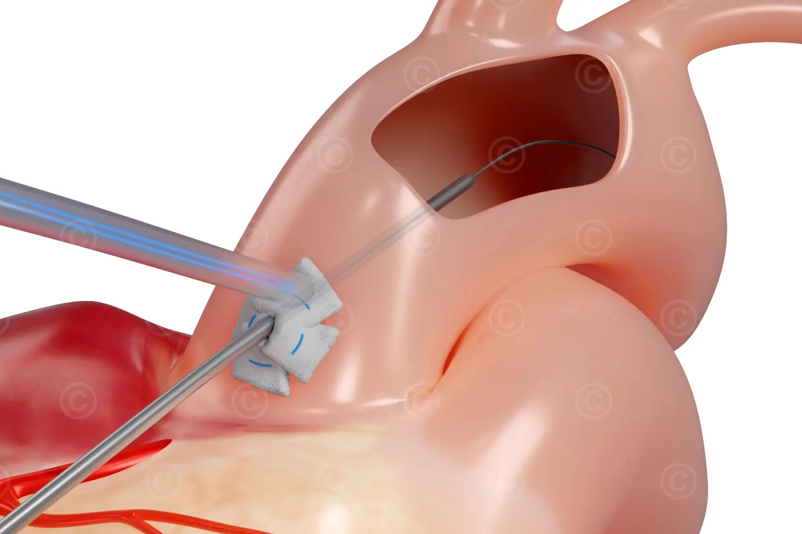
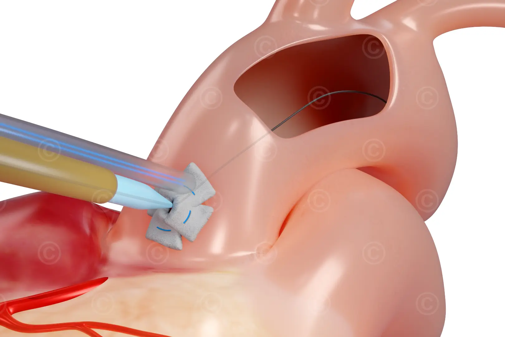
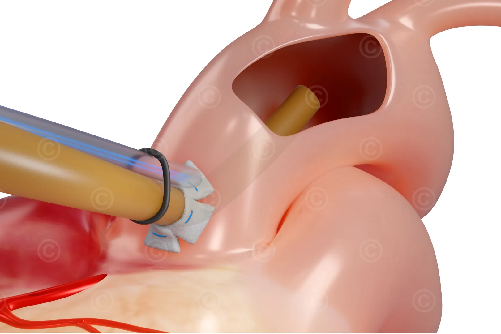
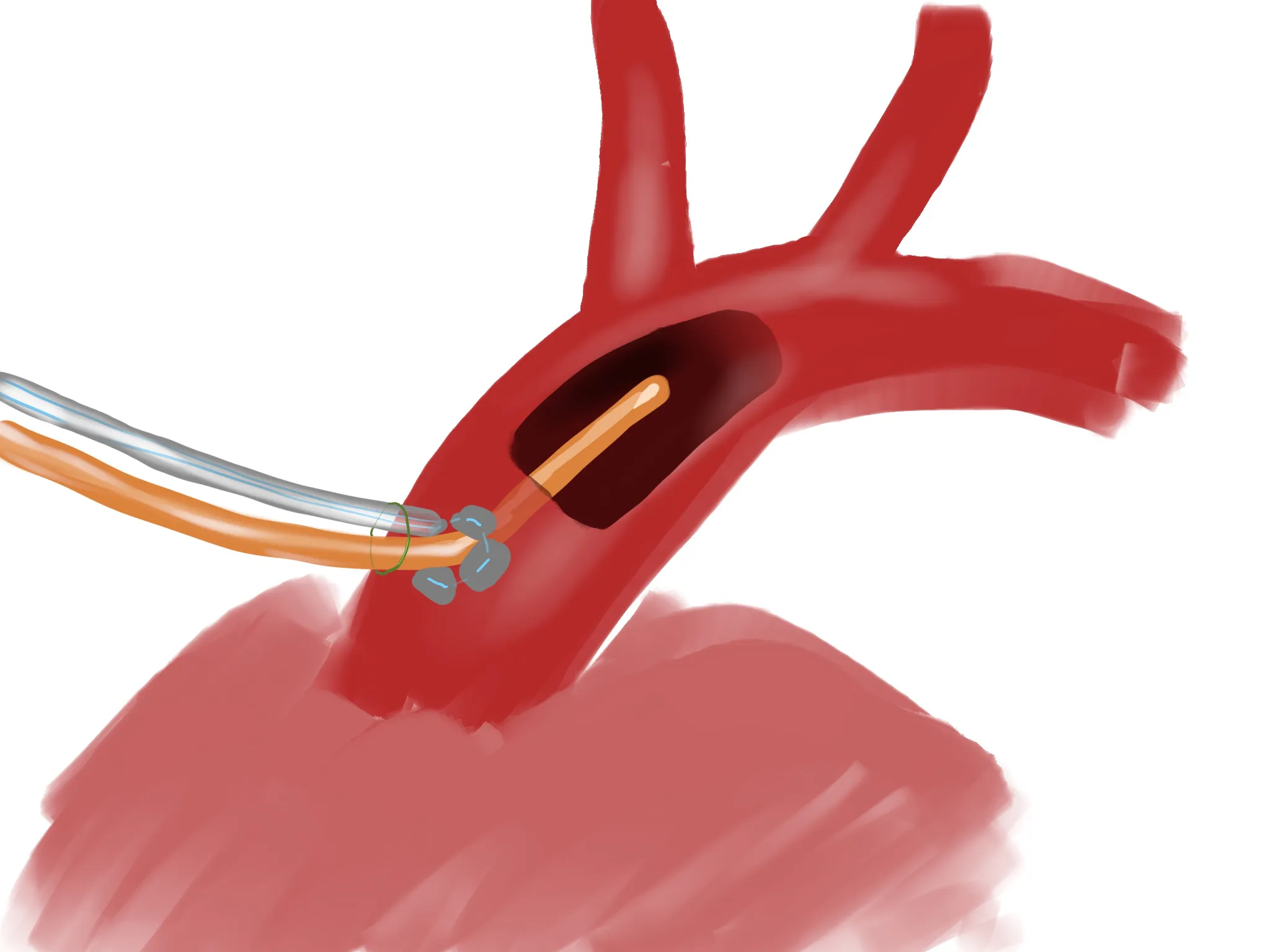
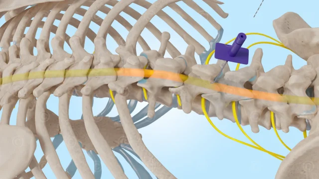
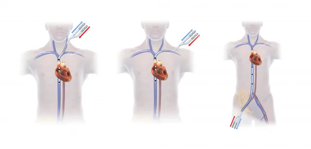
MRI-compatible catheters and instruments were also used for aortic puncture and cannulation. To minimize the risk of vascular injury and bleeding, the Seldinger technique was used for aortic cannulation (puncture with a needle, advancement of a guidewire, and placement of the catheter over the wire).
Study published April 27th, 2026 in “Journal of Cardiovascular Development and Disease“
Project details Illustrations for study on heart surgery with MRI
Content: 5 illustrations
Use: Publication
Specs: ca. DIN A4
Client: Vascular Surgery, Hirslanden / Clinic for Cardiac Surgery, University Hospital Zurich
The rights to use the images shown here belong to the client; use of the images shown here is not permitted. The images are protected by watermarks.