Background:
When evaluating scoliosis, it is important to be able to assess skeletal maturity in adolescents. The lower the level of maturity, the more growth can still be expected—and thus a potential increase in the curvature.
Project description:
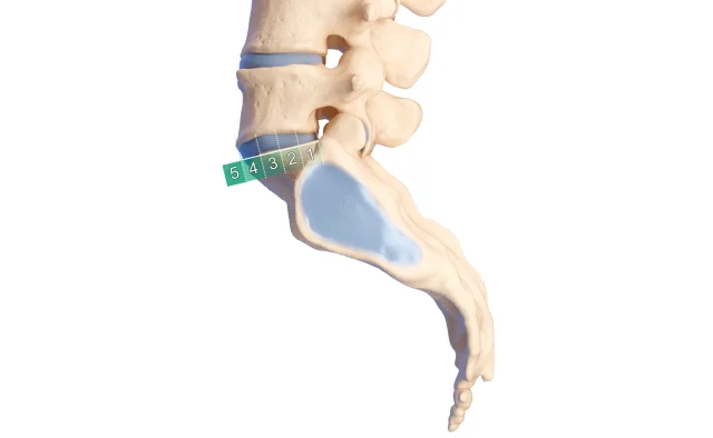
For an educational app developed by Aesculap AG, we created a series of illustrations on anatomy and spinal disorders. The Risser sign (Risser scale) is a radiological measure used to determine skeletal maturity and, consequently, the remaining growth potential of an adolescent. Using an X-ray of the pelvis, the degree of ossification of the apophyses (bone protrusions) of the iliac crest is assessed. During skeletal maturation, ossification does not occur simultaneously but progresses from the frontal/lateral to the medial (from the anterior superior iliac spine toward the sacrum). This area is divided into four segments for the Risser sign. The more advanced the ossification is in these segments, the more mature the skeleton is.
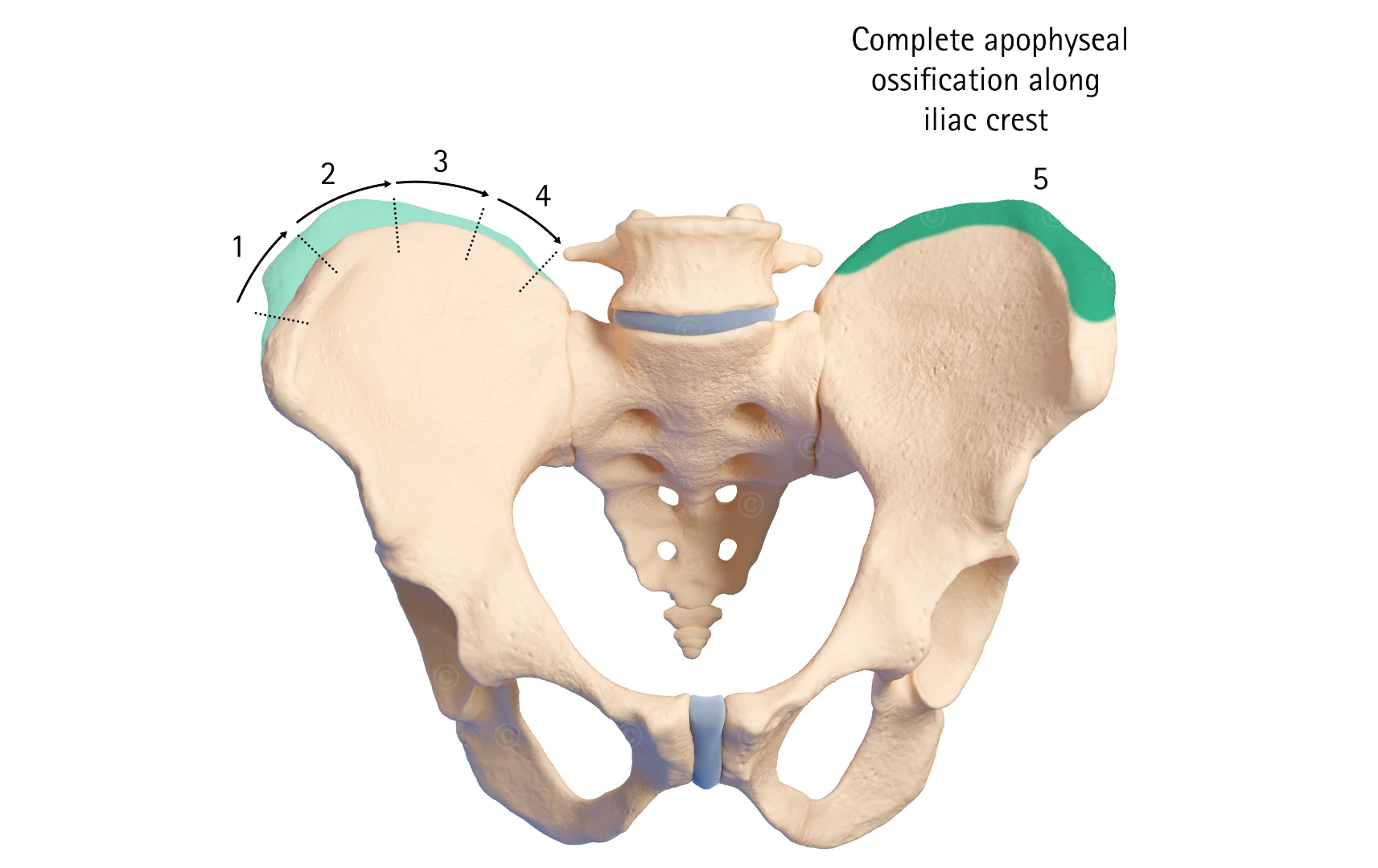
In our illustration, the sections corresponding to Risser stages I–IV are shown in light green on the left side of the image. On the right side of the image, the apophysis has completely fused with the ilium (dark green), indicating the end of growth.
Project details illustration Risser sign and skeletal maturity
Content: 1 Illustration
Use: e-Learning
Specs: ca. 4K
Client: Aesculap AG / B. Braun
The rights to use the images shown here belong to the client; use of the images shown here is not permitted. The images are protected by watermarks.