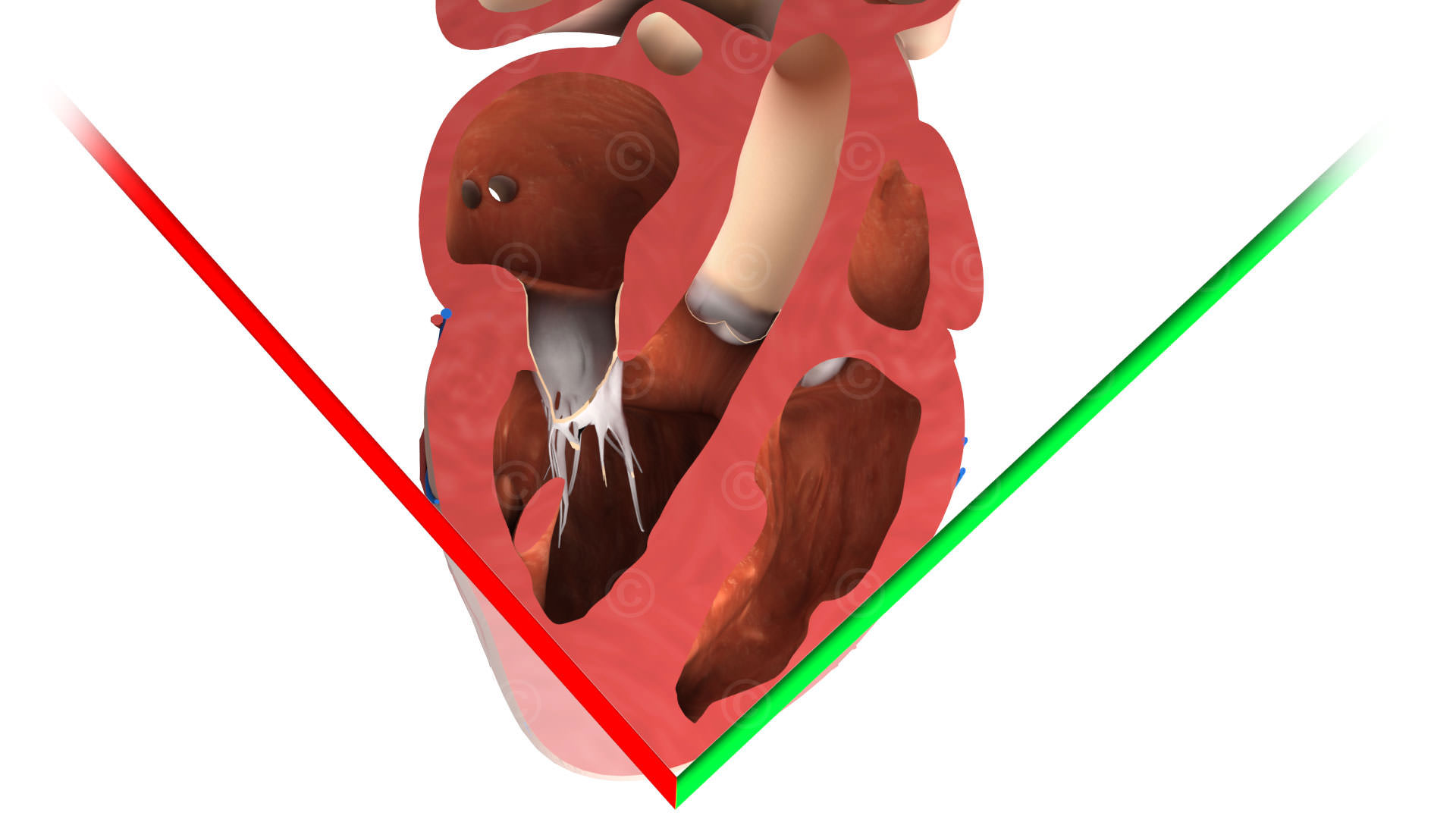
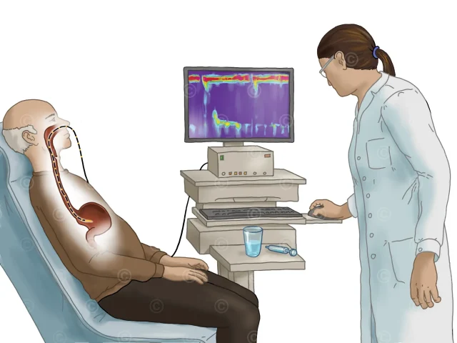
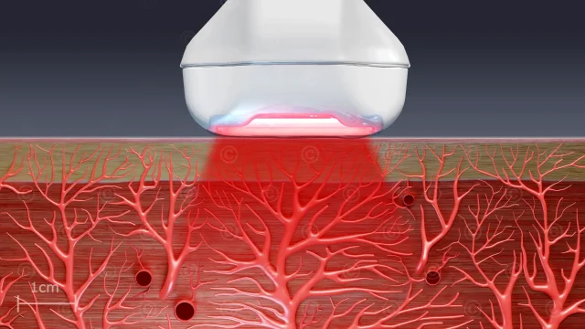
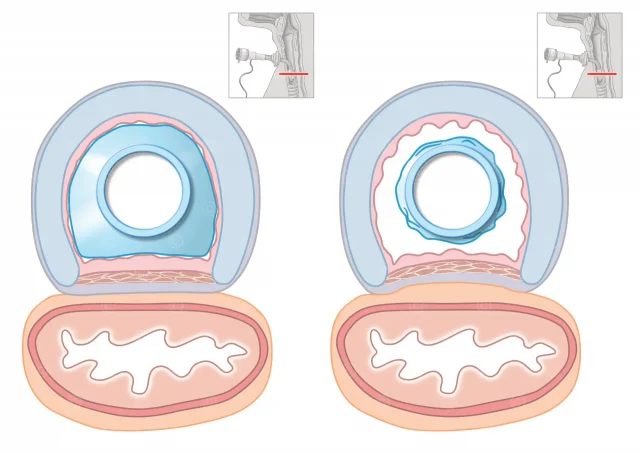
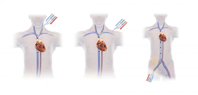
Illustrations of standard cuts of the heart with a transesophegal echo (TEE) for training and education. An ultrasound head is inserted into the esophagus and allows a sonography of different areas of the heart.
In addition to the illustrations, 3D PDFs of the cuts were created, which allow a better spatial coverage of the cutting planes. You can find more information in the article 3D-PDFs in der Sonografie (german).
Project details:
Content: 28 Illustration of the standard level in medical diagnostics – 29 interactive 3D-PDFs
Utilization: Training, educations, lectures
Specifications: Resolution 1920*1080 Pixel
Client: Robert Bosch Krankenhaus
The rights of use of the illustrations shown are with the respective clients.