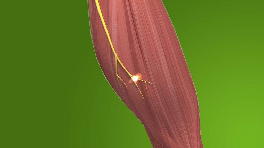
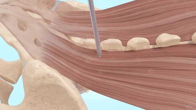
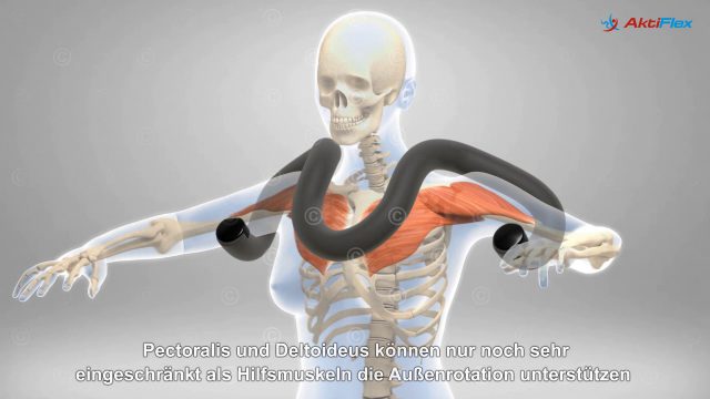
Design of a 3D animation on the anatomical structure of skeletal muscles, also called striated muscles, for the IST Study Institute for use in training and education. Skeletal muscles are responsible for the body’s ability to move and are formed of various elements.
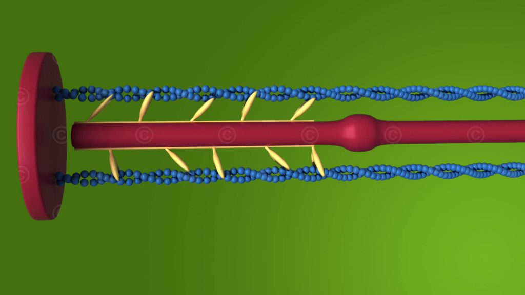
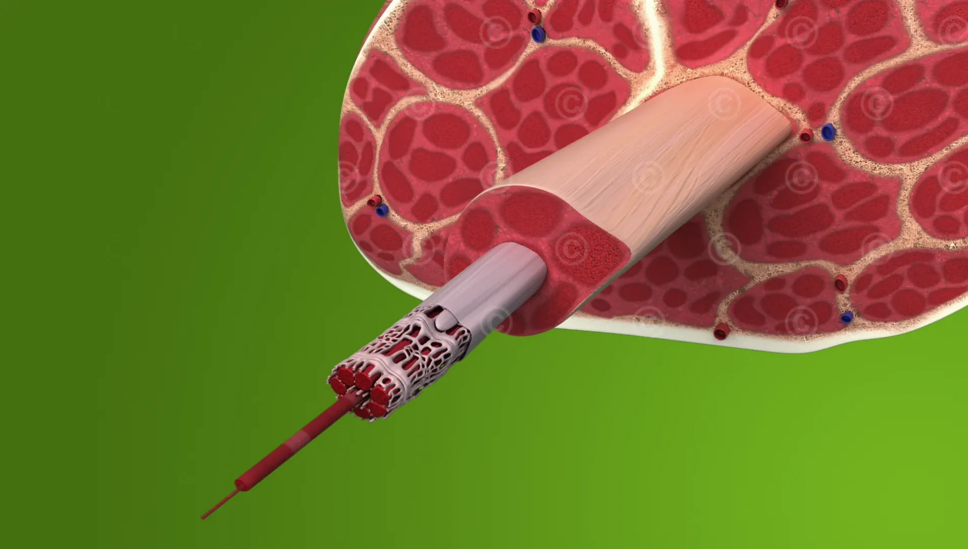
A muscle consists of a series of muscle fibers. These long, cylindrical cells contract when stimulated, causing muscle contraction. Muscle fibers, in turn, contain hundreds of myofibrils, which are divided into elongated sections called sarcomeres. Sarcomeres are the functional units within myofibrils. As the myofilaments actin and myosin slide into each other, the sarcomeres shorten (contract) and so do the entire myofibrils.
Please activate subtitles in the player for a description – Rights of use of the visual material: IST-Studieninstitut – Music: Kevin McLeod – Impact prelude – License: Creative Commons (CC BY 3.0)
Project details:
Content: 1 Animation
Utilization: Lectures, trainings, online courses
Specifications: Full HD – Resoulution 1920*1080 pixel
Client: IST-Studieninstitut
The rights of use of the illustrations shown are with the respective clients.
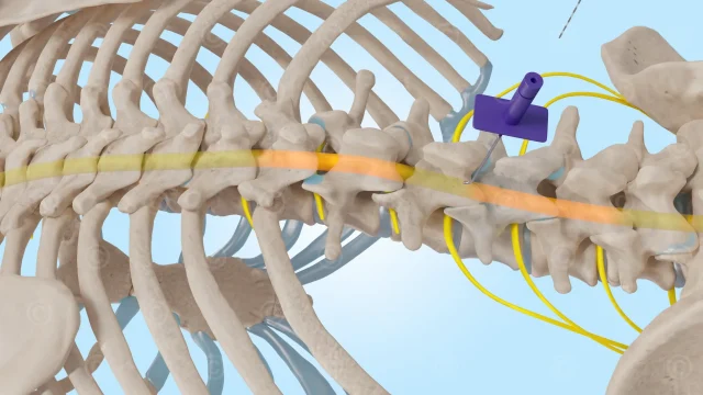
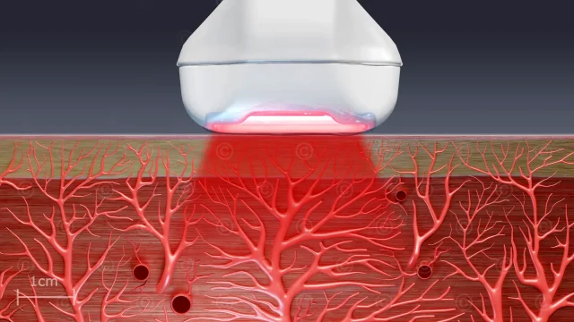
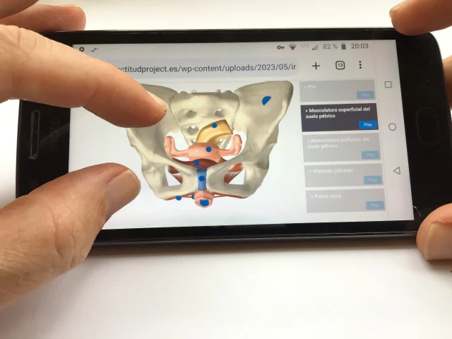
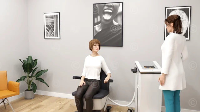
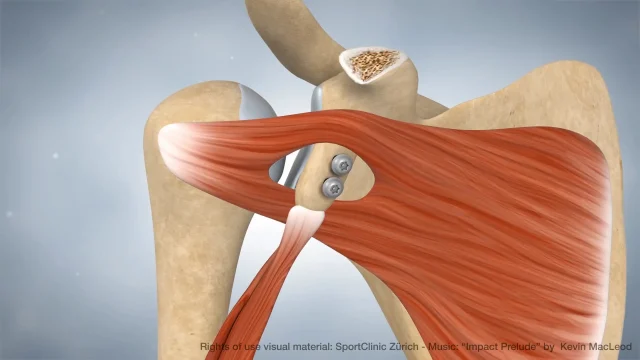
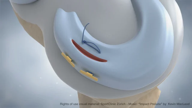
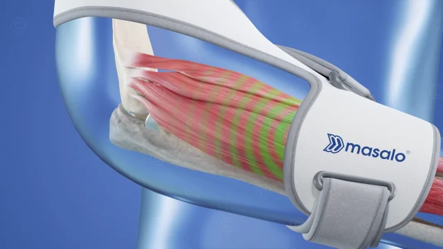
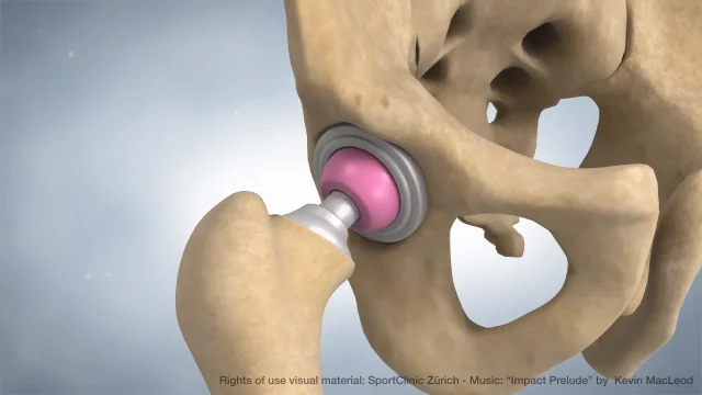
Screenshots Animation: